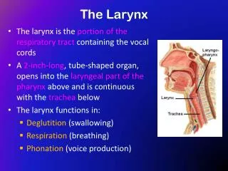
The larynx
The larynx. The Larynx. The larynx constitutes the organ of phonation and forms a protective sphincter to the respiratory system. It lies in the median part of the front of the neck, opposite the 3 rd , 4 th , 5 th and 6 th cervical vertebrae.

The larynx
E N D
Presentation Transcript
The Larynx • The larynx constitutes the organ of phonation and forms a protective sphincter to the respiratory system. • It lies in the median part of the front of the neck, opposite the 3rd, 4th, 5th and 6thcervical vertebrae. • It extends from the upper border of epiglottis to the lower border of cricoid cartilage. • Its upper end opens into the laryngopharynx by the laryngeal inlet. • Its lower end is continuous with the trachea at the level of the sixth cervical vertebra.
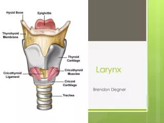
The Larynx • The larynx is formed by a cartilaginous skeleton attached together by joints and ligaments and moved by muscles and all are covered by mucous membrane. • Laryngeal cartilages: • Single: • Thyroid cartilage. • Cricoid cartilage. • Epiglottis cartilage. • Paired: • Arytenoid cartilages. • Corniculate cartilages. • Cuneiform cartilages.
Thyroid Cartilage • The largest laryngeal cartilage. • Formed of 2 quadrilateral laminae which are fused anteriorly. • Posteriorly, the 2 laminae are separated by a wide gap • The anterior border forms a median projection, the laryngeal prominence. • The laminae present a median V-shaped notch, the superior thyroid notch. • The posterior borders form upward and downward projections, superior and inferior horns. • The outer surface of each lamina shows an oblique ridge, the oblique line. • The oblique line extends between two tubercles, the superior and inferior thyroid tubercles.
Cricoid Cartilage • Smaller, but thicker than the thyroid cartilage. • Lies below and behind the thyroid cartilage. • It has the shape of a signet ring with a quadrilateral lamina posteriorly and a narrow arch anteriorly. • Its lower border is horizontal. • Its upper border is sloping. Epiglottis Cartilage • A leaf-shaped lamella of elastic cartilage. • It projects upwards behind the tongue and hyoid bone. • Has an upper broad free end and a lower tapering end. • Anteriorly, it is connected to: • The root of the tongue, by the median and lateral glosso-epiglottic folds. • The hyoid bone, by the hyoepiglottic ligament. • The inner surface of the thyroid cartilage by the thyroepiglottic ligament.
Arytenoid Cartilage • A pyramidal-shaped cartilage, with an apex, base and 3 surfaces: • Posterior surface. • Anterolateral surface. • Medial surface. • The apex is directed upwards and articulates with the corniculate cartilage. • The base is directed downwards and articulates with the upper border of the lamina of cricoid cartilage. • The lateral angle of the base projects and forms the muscular process. • The anterior angle of the base projects to form the vocal process.
Corniculate Cartilage • A small cartilage on the apex of the arytenoid cartilage • Enclosed in the aryepiglottic fold forming the corniculate tubercle Cuneiform Cartilage • A small nodule enclosed in the aryepiglottic fold forming the cuneiform tubercle
Joints of the Larynx • Cricothyroid joints: • One on each side. • A synovial joint between the inferior horn of the thyroid cartilage and the side of the cricoid cartilage. • Cricoarytenoid joints: • One on each side. • A synovial joint between the base of arytenoid cartilage and upper border of the lamina of cricoid cartilage. • Arycorniculate joints: • One on each side. • A joint between the apex of the arytenoid cartilage and the corniculate cartilage.
Membranes and ligaments • Thyrohyoid membrane and ligaments. • Fibroelastic membrane. • Quadrangular membrane. • Vestibular ligament. • Cricothyroid ligament (conus elasticus). • Vocal ligament. • Hyoepiglottic ligament. • Thyroepiglottic ligament. • Cricotracheal ligament.
Thyrohyoid membrane • Extends from the upper border of thyroid cartilage to the upper border of posterior surface of hyoid bone • Separated from the posterior surface of hyoid bone by the hyoid bursa. • The median part of the membrane is thickened to form the median thyrohyoid ligament. • The posterior border is thickened to form the lateral thyrohyoid ligament. • The membrane is pierced by the superior laryngeal vessels and the internal laryngeal nerve. • The lateral thyrohyoid ligament contains a cartilage nodule called the cartilago triticea.
Fibroelastic membrane • Its upper part is called quadrangular membrane and its lower parts is called cricothyroid ligament (conus elasticus). • The quadrangular membrane: • It extends between the lateral border of the epiglottis and the arytenoid cartilage. • Its lower free border forms the vestibular ligament. • The cricothyroid ligament (conus elasticus): • An elastic band which lies below and on the inner aspect of the thyroid cartilage. • It is connected to thyroid cartilage, cricoid cartilage and arytenoid cartilage. • Two parts of the ligament can be recognized:
Median cricothyroid ligament: • the anterior thickened part of the ligament which connects the adjacent borders of the cricoid and thyroid cartilages. • The ligament is overlapped by the cricothyroid muscle. • Lateral cricothyroid ligament (crico-vocal membrane) • this ligament is attached inferiorly to the upper border of cricoid cartilage • superiorly, it has 2 attachments: • to the inner surface of thyroid cartilage, anteriorly. • to the vocal process of arytenoid cartilage, posteriorly. • The free upper border of the crico-vocal membrane is called the Vocal Ligament. • So, the vocal ligament is attached to the inner surface of thyroid cartilage (anteriorly) and to the vocal process of the arytenoid cartilage (posteriorly).
Other ligaments • Hyo-epiglottic ligament: connects the anterior surface of epiglottis to the hyoid bone • Thyro-epiglottic ligament: connects the lower end of the epiglottis to the inner surface of thyroid cartilage • Crico-tracheal ligament: connects the lower border of the cricoid cartilage to the first ring of trachea • Ligaments of the joints.
Laryngeal Inlet • The opening of communication between the pharynx and larynx. • It is directed upwards and backwards • Boundaries: • Upper border of epiglottisanteriorly. • Aryepiglottic folds on both sides. • Mucous membrane between the arytenoid cartilages posteriorly. • Aryepiglottic fold: • Is a fold of mucosa extending between the apex of arytenoid cartilage and the side of epiglottis. • It encloses the aryepiglottic muscle, cuneiform and corniculate cartilages.
Laryngeal Cavity • Vocal fold: • A fold of mucous membrane extending from the inner surface of thyroid cartilageto the vocal process of arytenoid cartilage. • The folds enclose the vocal ligaments (upper border of lateral cricothyroid ligaments). • The fissure between the 2 vocal folds is called the rima glottidis. • Vestibular fold: • A fold of mucous membrane extending from the inner surface of thyroid cartilage to the anterolateral surface of arytenoid cartilage. • It encloses a fibrous band called the vestibular ligament. • Sinus of the larynx: • A recess between the vestibular and vocal folds • Vestibule of the larynx: • The part of the cavity between the inlet and the vestibular folds
Muscles of the larynx • Extrinsic muscle: • Elevators: • Digastric, stylohyoid, mylohyoid, geniohyoid, stylopharyngeus, salpingopharyngeus and palatopharyngeus. • Depressors: • Sternothyroid, sternohyoid, omohyoid muscles and the elastic recoil of the trachea. • Intrinsic muscles: • Acting on the laryngeal inlet: • Narrowing: oblique arytenoid and aryepiglottic muscles. • Widening: thyroepiglottic muscle. • Acting on the vocal cords: • Sphincteric action: • Adduction: lateral cricoarytenoid & transverse arytenoid. • Abduction: posterior cricoarytenoid. • Change in length and tension: • Elongation and tension: cricothyroid. • Shortening and relaxation: thyroarytenoid and vocalis.
Oblique arytenoid Muscle • Origin: from the back of the muscular process of the arytenoid cartilage. • Insertion: into the apex of the opposite arytenoid cartilage. • Nerve supply: recurrent laryngeal nerve (branch of vagus nerve. • Action: narrowing of the laryngeal inlet by bringing the two aryepiglottic folds together.
Aryepiglottic Muscle • Origin: from the apex of the arytenoid cartilage as a continuation of the oblique arytenoid muscle. • Insertion: into the side of the epiglottis cartilage. • Nerve supply: recurrent laryngeal nerve (branch of vagus nerve. • Action: narrowing of the laryngeal inlet by shortening the aryepiglottic fold.
Thyroepiglottic Muscle • Origin: from the inner surface of the thyroid cartilage. • Insertion: into the side of the epiglottis cartilage. • Nerve supply: recurrent laryngeal nerve (branch of vagus nerve. • Action: widening of the laryngeal inlet.
Lateral Cricoarytenoid Muscle • Origin: from the upper border of the arch of cricoid cartilage. • Insertion: into the muscular process of the arytenoid cartilage. • Nerve supply: recurrent laryngeal nerve (branch of vagus nerve. • Action: Adduction of the vocal cords by rotation of the arytenoid cartilage.
Transverse Arytenoid Muscle • Origin: from the back and medial surface of the arytenoid cartilage. • Insertion: into the back and medial surface of the opposite arytenoid cartilage. • Nerve supply: recurrent laryngeal nerve (branch of vagus nerve. • Action: Adduction of the vocal cords by approximating the arytenoid cartilages.
Posterior Cricoarytenoid Muscle • Origin: from the posterior surface of the lamina of the cricoid cartilage. • Insertion: into the muscular process of the arytenoid cartilage. • Nerve supply: recurrent laryngeal nerve (branch of vagus nerve. • Action: Abduction of the vocal cords by rotating the arytenoid cartilage.
Thyroarytenoid Muscle • Origin: from the inner surface of thyroid cartilage lateral to the cricothyroid ligament. • Insertion: into the antero-lateral surface of arytenoid cartilage. • Nerve supply: recurrent laryngeal nerve (branch of vagus nerve). • Action: shortening and relaxation of the vocal fold.
Vocalis Muscle • Origin: deep fibers of thyroarytenoid. • Insertion: into the vocal process of arytenoid cartilage and vocal ligament. • Nerve supply: recurrent laryngeal nerve (branch of vagus nerve. • Action: shortening and relaxation of the vocal fold.
Nerve Supply of the Larynx • Superior laryngeal nerve (from vagus nerve) divides into: • External laryngeal nerve (motor): supplies the cricothyroid muscle. • Internal laryngeal nerve (sensory): supplies the mucous membraneabove the level of the vocal folds. • Recurrent laryngeal nerve (from vagus nerve) divides into motor and sensory branches: • The motor branch supplies ALL intrinsic muscles of the larynx (except cricothyroid muscle). • The sensory branch supplies the mucous membrane below the level of the vocal folds.
Blood supply of the larynx • Arterial supply of the larynx: • Superior laryngeal artery (from superior thyroid artery from external carotid artery). • Inferior laryngeal artery (from inferior thyroid artery from thyrocervical trunk from 1st part of subclavian artery). Lymph drainage of the larynx To the deep cervical lymph nodes.
Clinical Points of the Larynx • Injury of the external laryngeal nerve: • Paralysis of the cricothyroid muscle which affects the voice (weakness) with no effect on air pathway. • Injury of the recurrent laryngeal nerve: • Unilateral complete section: the affected vocal cord will be between adduction and abduction (cadaveric position). The other cord will compensate and crosses to the other side and this will not affect the voice or respiration
Clinical Points of the Larynx • Bilateral complete section: both vocal cords will be between adduction and abduction (cadaveric position). The voice will be lost and the respiration is slightly affected. • Unilateral partial section: the affected side will show paralysis of the abductor muscle (posterior cricoarytenoid), the affected vocal cord will stay in adduction position, the other cord will compensate.
Clinical Points of the Larynx • Bilateral partial section: a severe condition because both vocal cords will be paralyzed in adducted position). The patient will suffer from stridor and asphyxia and tracheostomy or cricothyriodotomy is life saving process.