The Larynx
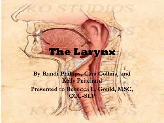
The Larynx. The larynx is the portion of the respiratory tract containing the vocal cords A 2-inch-long , tube-shaped organ, opens into the laryngeal part of the pharynx above and is continuous with the trachea below The larynx functions in: Deglutition (swallowing)

The Larynx
E N D
Presentation Transcript
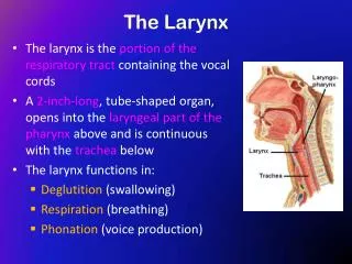
The Larynx • The larynx is the portion of the respiratory tractcontaining the vocal cords • A2-inch-long,tube-shaped organ, opens into the laryngeal part of the pharynxabove and is continuous with thetracheabelow • The larynx functions in: • Deglutition (swallowing) • Respiration (breathing) • Phonation (voice production)
The Larynx: Important Relations • The larynx related to major critical structures: • Carotid arteries , jugular veins, and vagus nerve • Superior and inferior thyroid arteries • Superior and recurrent laryngeal nerves
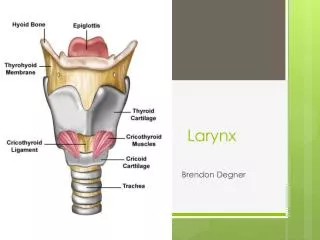
Structure • The larynx consists of four basic components: • Acartilaginous skeleton • Membranes and ligaments • Intrinsic and extrinsic muscles • Mucosal lining
The Cartilages • The cartilaginous skeleton is comprised of : • Single Cartilages: • Thyroid • Cricoid • Epiglottis • Paired Cartilages: • Arytenoid • Corniculate • Cuneiform
All the cartilages, except the epiglottis, are of hyaline type. Epiglottis is formed of elastic cartilage The cartilages are: Connected by joints, membranes & ligaments Moved by muscles
Thyroid Cartilage • Has two laminae, which meet in the midline and form a prominent angle, called laryngeal prominence (Adam’s apple) and the superior thyroid notch at the rostral margin of the • The posterior border of each lamina forms superior & inferior cornu (horns) • Outer surface of each lamina shows an oblique line which gives attachment to thyrohyoid, sternothyroid & inferior constrictor of the pharynx • Thesuperior border gives attachment to the thyrohyoid membrane superior cornu Oblique line inferior cornu
Cricoid Cartilage • Lies below the thyroid cartilage • Forms a complete ring • Has a narrow anterior arch & a broad posterior lamina • Has an articular facet on its: • Lateral surface for articulation with inferior cornu of the thyroid cartilage (a synovial joint) • Upper border for articulation with base of arytenoid cartilage (a synovial joint)
Arytenoid Cartilages • Small, pyramidal in shape • Situated at the back of the larynx Has: • Abase articulating with the upper border of the cricoid cartilage • Anapex supporting the corniculate cartilage • Avocal process projecting forward, gives attachment to the vocal ligament • Amuscular process projecting laterally, gives attachment to muscles
Corniculate & Cuneiform Cartilages E Corniculate Cartilages • Small nodules • Articulate with the apices of arytenoid cartilages Cuneiform Cartilages • Small rod shaped, placed in each aryepiglottic fold, producing a small elevation • Do not articulate with any other cartilage Serve as support for the ary-epiglottic fold VF CU CO
Epiglottis • Leaf shaped, situated behind the root of the tongue • Connected: • In front to the body of hyoid bone by the hyoepiglottic ligament • By its stalk to the back of thyroid cartilage by the thyroepiglottic ligament • Upper edge is free. • Laterally gives attachment to aryepiglottic fold • Anteriorly mucosa is reflected onto the tongue forming three glossoepiglottic folds & valleculae
Membranes & Ligaments • Thyrohoid membrane, median & lateral thyrohoid ligaments • Median cricothyroid ligament • Cricotracheal membrane • Hyoepiglottic ligament • Thyroepiglottic ligament
Quadrangular membrane: • Extends between the epiglottis and the arytenoid cartilages • Its lower free margin forms the vestibular ligament that lies within the vestibular fold • Cricothyroid membrane (conus elasticus): • Lower margin is attached to upper border of cricoid cartilage • Upper free margin forms vocal ligament that is attached anteriorly to deep surface of thyroid cartilage & posteriorly to the vocal process of arytenoid cartilage
Laryngeal Inlet • Faces backward and upward and opens into the laryngeal part of the pharynx • The opening is bounded: • Anteriorly: by the upper margin of epiglottis • Posteriorly & below by arytenoid cartilages • Laterally by aryepiglottic folds E CU CO AEF A
Laryngeal Cavity • Extends from laryngeal inlet to lower border of the cricoid cartilage • Narrow in the region of the vestibular folds (rima vestibuli) • Narrowest in the region of the vocal folds (rima glottidis) Rima vestibuli Rima glottidis
Laryngeal Cavity cont’d • Divided into three parts: • Supraglottic part, the part above the vestibular folds, is called the vestibule • The part between the vestibular & the vocal folds, is called the ventricle • Infraglottic part, the part below the vocal folds A B C
Vestibular Part: • Extends from the inlet to the vestibular fold • Below it becomes narrow as the vestibular folds project medially. • Each vestibular fold contains vestibular ligament, the lower free margin of the quadrangular membrane stretching from thyroid cartilage to the arytenoid cartilage • Lower Part: • Extends from vocal folds to lower border of cricoid cartilage • Walls formed by the inner surface of the cricothyroid ligament and the cricoid cartilage
Middle Part • Extend from vestibular folds to the vocal folds • Laterally a small recess between the vestibular fold & the vocal fold is called the sinus of the larynx, which may extend upwards between vestibular fold and the thyroid cartilage as saccule of the larynx
Mucous Membrane • The cavity is lined with ciliated columnar epithelium • The surface of vocal folds, because of exposure to continuous trauma during phonation, is covered with stratified squamous epithelium • Contains many mucous glands, more numerous in the saccule (for lubrication of vocal folds) Muscles: Divided into two groups: • Extrinsic muscles: divided into two groups • Elevators of the larynx • Depressors of the larynx • Intrinsic muscles: divided into two groups • Muscles controlling the laryngeal inlet • Muscles controlling the movements of the vocal cords
Elevators of the Pharynx • The Suprahyoid Muscles • Digastric • Stylohyoid • Mylohyoid • Geniohyoid • The Longitudinal Muscles of the Pharynx • Stylopharyngeus • Salpingopharyngeus • Palatopharyngeus Depressors of the Pharynx: • The Infrahyoid Muscles • Sternohyoid • Sternothyroid • Omohyoid
Muscles Controlling the Laryngeal Inlet • Oblique arytenoid • Aryepiglottic muscle
Muscle Increasing the Length & Tension of the Vocal Cords • Cricothyroid: increases the distance between the angle of the thyroid cartilage & the vocal processes of the arytenoid cartilages, and results in increase in the length & tension of the vocal cords
Muscle decreasing the Length & Tension of Vocal Cords • Thyroarytenoid (vocalis): pulls the arytenoid cartilage forward toward the thyroid cartilage and thus shortens and relaxes the vocal cords
Movements of the Vocal Cords • Adduction • Abduction Glottis (space between folds) Folds closed (adducted) Folds open (abducted) (View from above)
Adductors of the Vocal Cords • Lateral cricoarytenoid • Transverse arytenoid
Abductor of the Vocal Cords • Posterior cricoarytenoid
Sphincteric Function of the Larynx There are two sphincters: • At the inlet: used only during swallowing • At the rima glottis: used in coughing and sneezing
Blood Supply & Lymph Drainage • Arteries: • Upper half: Superior laryngeal artery, branch of superior thyroid artery • Lower half: Inferior laryngeal artery, branch of inferior thyroid artery • Veins: • Accompany the corresponding arteries • Lymphatics: • The lymph vessels drain into the deep cervical lymph nodes
Nerve Supply • Sensory • Above the vocal cords: Internal laryngeal nerve, branch of the superior laryngeal branch of the vagus nerve • Below the vocal cords: Recurrent laryngeal nerve, branch of the vagus nerve • Motor • All intrinsic muscles, except cricothyroid, supplied by the recurrent laryngeal nerve • The cricothyroid muscle is supplied by the external laryngeal nerve,a branch of the superior laryngeal branch of vagus nerve
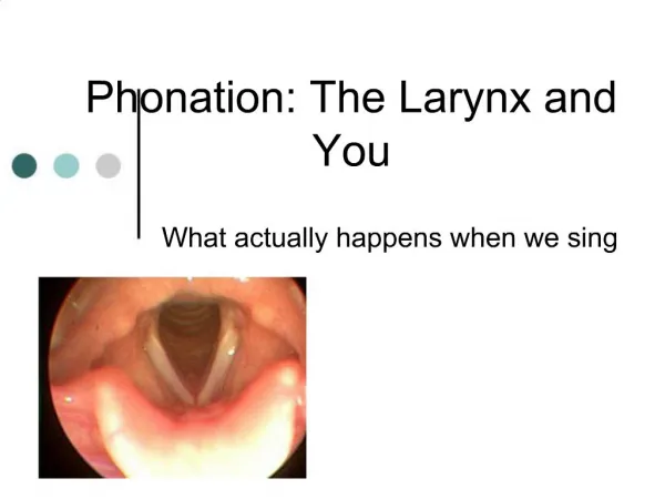
Production of Voice • The production of voice has three components: • The generation of sound:Sound production originates from the larynx as afundamental tone by the intermittent release of expired air between the adducted vocal cords resulting in their vibration. • The resonance of sound: This tone is modified by various resonating chambers (resonators) i.e. pharynx, mouth and paranasal sinuses. • The articulation of voice (speech production) : Finally converted to speech by the action of the mouth, nose, nasal cavity and throat, where the tongue, palate, cheek and lips are involved in articulation Parameters of Voice • Quality, Loudness, andPitch • Quality :depends on symmetrical vibration at the midline of the glottis • Loudness : is influenced by subglottic pressure, glottic resistance, transglottic air flow, and amplitude of vibration • Pitch : depends on the alterations in length and tension of vocal folds
Clinical Notes • Laryngitis • Edema of laryngeal mucosa • Laryngeal nerve lesions: • External laryngeal nerve • Unilateral • Bilateral • Recurrent laryngeal nerve C. Unilateralcomplete (of right nerve) D. Bilateralcomplete E. Unilateralpartial (of right nerve) F. Bilateralpartial The position of vocal cords