M ass S pectrometer(MS)
M ass S pectrometer(MS). 분자가 MS 내로 들어가면 분자는 이온화됨과 동시에 더 작은 이온들 (fragments) 로 쪼개진다 . 쪼개진 이온들은 그들의 질량 / 전 하 (m/z ) 비에 따라 선택적으로 분리되어 이온수에 비례해 signal 을 만든다 . 이온들의 질량 / 전하비는 이온들의 생성된 양 (Abundance) 의 함수로 표시되어 mass spectrum 이 그려지며 , 이 mass spectrum 을 이용하여 미지성분의 정성확인을 할 수 있다 .

M ass S pectrometer(MS)
E N D
Presentation Transcript
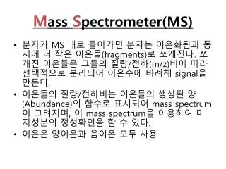
Mass Spectrometer(MS) • 분자가 MS 내로 들어가면 분자는 이온화됨과 동시에 더 작은 이온들(fragments)로 쪼개진다. 쪼개진 이온들은 그들의 질량/전하(m/z)비에 따라 선택적으로 분리되어 이온수에비례해 signal을 만든다. • 이온들의 질량/전하비는이온들의 생성된 양(Abundance)의 함수로 표시되어 mass spectrum이 그려지며, 이 mass spectrum을 이용하여 미지성분의 정성확인을 할 수 있다. • 이온은 양이온과 음이온 모두 사용
MS component • Sample inlet 시료도입장치로 시료를 MS내로 효율적으로 보내주는 역할을 한다 • Ion source 시료분자를 이온화시키고 더 작은 이온으로 쪼갠다. 생성된 이온들을 MS analyzer 쪽으로 이동시킨다 • Mass analyzer 이온들을 m/z ratio에 따라 선택적으로 분리시킨다 • Ion detector 이온 흐름을 그 양에 비례하게 전기적인 흐름으로 전환, 증폭시켜 signal을 생성한다 • Vacuum system MS 내 진공상태를 10-4 ~ 10-9Torr로 만들어 주어 최적의 상태로 분석이 진행될 수 있도록 한다 • Data System MS 내 각 구성성분들의 조절이 가능하며 시료분석과 동시에 데이터해석을 할 수 있는 곳이다
Ionizationmethod TypicalAnalytes SampleIntroduction MassRange MethodHighlights Electron Impact (EI) Relativelysmallvolatile GC orliquid/solidprobe to1,000Daltons Hard methodversatileprovidesstructure info Chemical Ionization (CI) Relativelysmallvolatile GC orliquid/solidprobe to1,000Daltons Soft methodmolecular ionpeak [M+H]+ Electrospray (ESI) PeptidesProteinsnonvolatile LiquidChromatographyor syringe to200,000Daltons Soft methodions oftenmultiplycharged Fast Atom Bombardment (FAB) CarbohydratesOrganometallicsPeptidesnonvolatile Sample mixedin viscousmatrix to6,000Daltons Soft methodbut harderthan ESI orMALDI Matrix Assisted Laser Desorption(MALDI) PeptidesProteinsNucleotides Sample mixedin solidmatrix to500,000Daltons Soft methodvery highmass Sample introduction / ionization method:
Ion source • Gaseous sample introduction - EI(electron ionization) - CI(chemical Ionization) • Liquid sample introduction - FAB(fast atom bombardment) - ESI(electrospray ionization)(soft ionization) • Solid sample introduction - MALDI(soft ionization) (matrix-assisted laser desorption/ionization)
Ion Source - EI(electron ionization) • 전자를 발생시키기 위한 filament가 가열되고 (+) 극판에 전압이 걸리면 가속된 전자들이 흐르고 그 속을 지나던 기체화된 sample은 전자와 충돌하여 에너지를 얻고 전자 하나를 잃어 분자이온 M+가 된다 • 이 방법은 큰 에너지를 사용하기 때문에 복잡한 spectrum을 이루며 분자 이온을 얻기 힘들다 • M + e-⇒ M+ + 2e-
Ion Source – CI(chemical Ionization) • 가열된 filament에서 발생, 가속된 전자는 106정도로 많은 reagent gas와 충돌하여 이들을 이온화시키고 이 reagent gas ion은 sample gas와 충돌 • 이 sample gas는 fragmentation 되기도 하고 때로는 reagent gas ion 과 complex를 이루기도 한다. • 매우 낮은 에너지로 충돌하기 때문에EI 보다 수백배 이상 많은 수의 분자 이온을 만들어 내기 때문에 분자량 확인에 많이 쓰인다. • R(CH4) + e-⇒ R+ + 2e- R+ + M ⇒ M1+ + N1 M1+ ⇒ M2+ + N2
Ion Source: ESI Electrospray ionization(ESI) • 용액 상태의 시료를 이온화(LC-MS) • 기존의 방법으로는 얻기 힘들었던 intact 상태의 peptide나 단백질을 이온화 • 한 개 이상의 전하를 띤 이온을 생성
Ion Source: ESI • 시료용액이 고전압이 걸려 있는 capillary를 통과하면서 분무되어 전하를 많이 띤 droplet이 생성됨 • dropolet이 capillary에서 orifice를 지나면서 inert gas(or heat)에 의해 desolvation • Desolvation과정에서 ion의 charge는 더 증가하고 “Coulombicexplosion”에 의해 droplet의 ion이 gas phase로 된다. • sample에 가해지는 충격이 약하며, multiple charge를 가진 peptide이온이 생긴다.
+ + Laser + m m m a + a a m m m m + m a a a m matrix + analyte m Sample support Ion Source: MALDI Matrix Assisted Laser Desorption Ionization(MALDI)
Why MALDI? -Less sensitive to salts -Lower PRACTICAL detection limits -Easier to interpret spectra(less multiple charges) -Quick and easy -Higher mass detection -Higher Throughput(1000>samples per hour)
Biomolecule Analysis 과거에는? • Electrophoresis, chromatography, ultracentrifugation • Not very precise MS이용하면? • Proteins, oligonucleotides, oligosaccharides, lipids • Detect modifications and sequences
Biomolecule Analysis • Mass is one of first measurements to characterize biopolymers • Up to 1970s, had to use–Electrophoresis–Chromatography–Ultracentrifugation–Not very precise (10 –100% relative error!!) • May use MS on most biomolecules–Proteins–Oligonucleotides–Oligosaccharides–Lipids • May detect modifications and sequences–Post translational and other
Peptide Mass Fingerprinting • Analytical technique for protein identification (protein sequence) • Unknown protein of interest cleaved into peptide by protease • Collection of peptides resulting from this cleavage comprise a unique identifier of the unknown protein • Mass measured with MALDI-TOF and ESI-TOF • in silico compared to the genome
Computer programs translate the known genome of the organism into proteins • Theoretically cut the proteins into peptides with the same protease (ex.Trypsin: K or R) • Calculate the absolute masses of the peptides from each protein • the masses of the peptides of the unknown protein vs the theoretical peptide masses of each protein encoded in the genome • Results statistically analyzed to find the best match
Trypsin Digest Cut out 2D-Gel Spot Protein Peptides
Peptide Mass Fingerprinting N K K K R K K R Trypsin K K K K R K R R Protein R R C N K K Tryptic peptide mixture. Masses measured by MS. Every peptide has a basic C-terminus. R C A protein can be identified in a database by matching masses of a subset of the tryptic peptides against calculated values.
peptide fragments intact protein enzyme MEMEKEFEQIDKSGSWAAIYQDIRHEASDFPCRVAKLPKNKNRNRYRDVS PFDHSRIKLHQEDNDYINASLIKMEEAQRSYILTQGPLPNTCGHFWEMVW EQKSRGVVMLNRVMEKGSLKCAQYWPQKEEKEMIFEDTNLKLTLISEDIK SYYTVRQLELENLTTQETREILHFHYTTWPDFGVPESPASFLNFLFKVRE SGSLSPEHGPVVVHCSAGIGRSGTFCLADTCLLLMDKRKDPSSVDIKKVL LEMRKFRMGLIQTADQLRFSYLAVIEGAKFIMGDSSVQDQWKELSHEDLE PPPEHIPPPPRPPKRILEPHNGKCREFFPNHQWVKEETQEDKDCPIKEEK GSPLNAAPYGIESMSQDTEVRSRVVGGSLRGAQAASPAKGEPSLPEKDED HALSYWKPFLVNMCVATVLTAGAYLCYRFLFNSNT
In Silico Digestion 812.6 1432.3 3127.1 996.8 702.4 164.9 2748.2 848.3 1272.7 493.2 882.6 2978.3 364.1 948.9 3128.8 3514.2 2837.1 263.9 147.4 1429.7 199.6 142.3 640.8 MS Peptide Mass Fingerprinting 2D-Gel Database “Spot removal” In Gel Digestion 848.1 1272.5 492.6 883.2 2978.9 Is identical to
Deduction of Full Amino Acid Sequence of a Protein by Overlapping the Sequences Obtained from individual Peptides
Edman Degradation Sequentially Removes One Residue at a Time from the Amino End of a Peptide up to 50 times Each round can be complete within 1 hr and the Edman degradation can be repeated up to 50 cycles in Practice.
Measured peptide mass 와 sequence가 맞지 않는 경우 • The additional masses are due to posttranslational or artifactual modifications or post-translational processing • Unspecific proteolysis had occurred or contaminating protease was present • Protein was part of a mixture of ‘contaminating’ proteins
Post Translational Modifications(PTM’s) • PTM’s are very important in signaling as well as metabolic pathways (e.g. phosphorylation) • Often we want to know not only which modification a protein has undergone, but exactly where in the sequence the modification lies. • Many of the search engines allow for “variable” modifications, but very few at one time (combinatorialy explosive) • There is great opportunity here for robust searches that find PTM’s reliably!
Phosphorylation site analysis strategies • Complication of phosphoprotein analysis - the frequently low stoichiometry of phosphorylation - the presence of multiple, differentially phosphorylated forms • In vitro analysis - scale up of protein by kinase reaction - comparison with 2D-PP maps of in vivo (confirmation of identity indirectly) - MS analysis
Detection and isolation of phosphoproteins • For the analysis of the site(s) of protein phosphorylation - purification of phosphoprotein - enzymatic or chemical fragmentation of the phosphoprotein - Isolation, separation, analysis of peptide • Isolation - separation of proteins by gel electrophoresis - fragmentation of the phosphoprotein band or spot - extraction of the generated phosphopeptide • More positive identification - 32 P radiolabelling : in vivo(32 PO4), in vitro([γ-32P]ATP) - western blotting : particularly tyrosine phosphorylated protein
Separation of phosphopeptides • 필요한 이유 - 농도를 농축하는 역할을 하여 S/N 비를 높임 - radiolabel의 activity를 이용하여 phosphopeptide의 상대적 또는 절대적인 양을 구할 수 있음 - separation에 의해 확보된 재현성으로 단백질의 phosphorylation 상태를 정량적으로 결정할 수 있음 - nonpeptide contaminants를 제거하여 적은 양의 phosphopeptide의 분석을 용이하게 함
Phosphopeptide separation techniques • By 2-dimensional phosphopeptide map • Reversed-phase HPLC • High-resolution gel electrophoresis • Immobilized metal affinity chromatogrphy(IMAC) • Phosphopeptide는 상대적, 절대적으로 적은 양 때문에 분석이 어려우므로 이러한 점을 극복할 수 있는 최적의 separation방법을 선택해야
Separation by 2D-PP • 1st dimension by electrophoresis on thin-layer cellulose plate + 2nd dimension by TLC on the same plate • information - radiolabelled spot 수 ⇒ phosphorylated sites 최대수 - radiolabelled spot의 intensity ⇒ peptide 들의 상대적인 phosphorylation 정도 - relative state between phosphopeptide • MS analysis after extraction from plate - protease양이 중요 • sensitive and reproducible by radiolabelling
Separation by RP-HPLC • Reproducible and simple • column으로 분리하고 radioactivity count로 fraction ⇒ count를 시간의 함수로 하여 radioactive fraction의 수를 알 수 있음 • 단점 - very hydrophilic phosphopeptide, very hydrophobic phosphopeptide의 분리가 어렵다 - 2D-PP보다 resolution이 낮다 - phosphopeptide will stick to metal surface • 장점 - ESI MS와 on line으로 연결하여 사용할 수 있다(LC-MS/MS) - isotope을 사용할 수 없는 인체 단백질 분석 가능
Separation by high-resolution electrophorsis and IMAC • High-resolution gel electrophoresis - 2-DE - 특정 phosphopeptide의 손실이 많지만 널리 보급되어 있어 사용하기 좋음 • IMAC - 같은 sequence를 갖는 nonphosphorylated peptide에 비하여 상대적으로 매우 적은 양의 phosphorylated peptide의 분석 어려움 - separation and enrichment 1) phosphopeptide와 metal(Fe3+ ,Ga3+)의 chelating 2) elution by phosphate or increased pH 3) acidic amino acid 도 enrichment 되는 단점
Determination of the type of phosphorylated amino acid • 이유 가능한 phosphorylated site의 수를 찾아냄으로써 polypeptide 내의 phosphorylated residues의 assignment를 쉽게 할 수 있음 • Technique 1) phosphoamino acid analysis - 32P-amino acid(hydrolysate of 32P-labeled phosphoprotein or phosphopeptide) ⇒ autoradiography - phosphoamino acid standard ⇒ ninhydrin staining - sample과 standard의 비교 분석(보통 1site/phosphopeptide) 2) phospho-amino acid-specific immunodetection - antibodies specific for particular phospho-amino acid - 상업적으로 antibody판매
Determination of the site of phosphorylation • Chemical phosphopeptide sequencing - phosphopeptide sequencing by step-wise chemical degradation(nonradioactive, radioactive methods) - analyzed as phenylthiohydantoyl derivatives - not available in very limited amount • Mass spectrometric analysis of phosphopeptides - phosphopeptide의 양이 1pmole이상이면 2D-PP map에서 extraction이 가능하고, MS로 분석이 가능 - two basic theme 1) chemical lability of the phosphate ester bonds 2) the detection of the mass added to a peptide (80u) - product ion scan in a tandem MS으로 phosphorylation site 확인 ⇒ phosphorylated amino acid type을 알고 있으면 더 용이
Mass scan for phosphopeptides analysis • In-source CID - identify phosphopeptides by observation of H2PO4-(97U), PO3-(79U) and PO2-(63U) - detect phosphopeptides in negative ion mode and then switch to positive ion mode • Neutral loss scan - positive ion mode with ESI in a TQ MS - Q1, Q3 are scanned over different m/z ranges - neutral loss of phosphoserine and phosphothreonine : 98
Mass scan for phosphopeptides analysis • Presursor ion scan - negative ion ESI(다시 positive ion mode로 변경) - Q1 : continous scan, Q2 : ion fragmentation Q3 : 79m/z(PO3-)를 잃은 ion 만 통과 • Product ion scanning - in-source CID, neutral loss and precursor ion scanning는 특수한 경우에만 phosphorylated residue를 identify - 상기 3가지 방법을 이용할 수 없는 경우 peptide fragment ion 전체에 대한 해석이 필요
Mass scan for phosphopeptides analysis • Post-source decay MALDI • Enzymatic and chemical dephosphorylation - MALDI-TOF로 phosphopeptide의 mass 측정 + phosphate를 제거 후 MALDI-TOF로 mass 측정 - nonphosphorylated peptide에서 phosphopeptide 를 확인하는데 쉽게 이용 - identification of phosphorylation sites using MS/MS
Emerging methods and future directions in phosphoprotien analysis • in vivo와 비교해서 in vitro로 시험하나 동일하다는 보장이 없음 ⇒ in vivo를 직접 • in vivo 32P-labeled protein을 충분히 얻는다는 것은 어려우므로 분석기기 감도를 높이는 것이 유리
Present and future challeges and opportunities • Protein identification and characterization has to be performed in a high-throughput manner, efficiently and with high accuracy and sensitivity • Robotic system • 2D-chromatography MS/MS