Urinary System Chapter 17
Urinary System Chapter 17. Objectives: 13.0 Identify structures and functions of the urinary system 13.1 Tracing the filtration of blood from the kidneys to the urethra 13.2 Recognizing diseases and disorders of the urinary system. Urinary System:.

Urinary System Chapter 17
E N D
Presentation Transcript
Urinary SystemChapter 17 Objectives: 13.0 Identify structures and functions of the urinary system 13.1 Tracing the filtration of blood from the kidneys to the urethra 13.2 Recognizing diseases and disorders of the urinary system
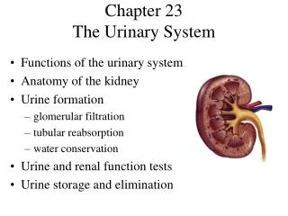
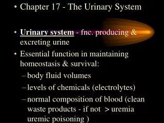
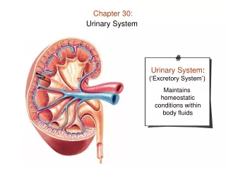
Urinary System: • removes salts and nitrogenous wastes from the blood. • helps maintain normal concentrations of water and electrolytes of bodily fluids • regulates pH and volume of bodily fluids • helps control RBC production and blood pressure
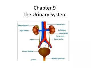
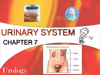
Organs of the Urinary System • Kidneys • Urinary bladder • They are connected by the ureters. • The urethra takes urine from the bladder to the outside of the body.
Kidneys • Located on either side of the vertebral column, on the posterior wall of the abdominal cavity. • Left is 1.5-2.0 cm higher than the right • Where do people feel kidney pain?
Kidney Structure • Lateral surface is convex; medial side is deeply concave. • Medial depression leads to hollow chamber called the renal sinus. • Entrance to sinus is called hilum, and it is also the passage for blood vessels, nerves, lymphatic vessels, and the ureter.
Kidney Structure, continued….. • Superior end of the ureter forms the renal pelvis (funnel-shaped sac inside the renal sinus) • Renal pelvis subdivides into major calyces (tubes, which divide into minor calyces. • Renal papillae project into the renal sinus.
Kidney Structure, continued….. • Two distinct regions in the kidneys: • Renal medulla (inner) • Composed of conical masses (renal pyramids) that appear striated • Renal cortex (outer) • Forms a shell around medulla • Projects into medulla between renal pyramids, forming renal columns
Kidney Functions • Regulate composition and volumes of extracellular fluids • Secrete hormone erythropoietin (???) • Role in activation of vitamin D • Help maintain blood pressure • Extracellular fluid volume • Secrete enzyme renin
Nephrons • The kidneys’ functional units • About 1 million in each kidney • Each nephron consists of: • Renal corpuscle • Renal tubule
Nephron, continued….. • Renal corpuscle composed of: • Glomerulus – tangled cluster of capillaries that filter fluid • Glomerular capsule – • sac-like structure surrounding the glomerulus • Located at the proximal end of the renal tubule • Receives the fluid filtered by the glomerulus
Nephron, continued….. • Renal tubule: • Transports fluid from the glomerular capsule to a minor calyx • Proximal convoluted tubule: • Dips down toward the renal pelvis and becomes the descending limb of the nephron loop (“loop of henle”) • Curves back up (ascending limb of the nephron loop) • Becomes coiled again (distal convoluted tubule)
Nephron, continued….. • Distal convoluted tubules from several nephrons will merge in the renal cortex to form a collecting duct • In the renal medulla, several collecting ducts will merge before emptying into a major calyx through an opening in a renal papilla.
Renal Blood Flow • Renal arteries branch off the _________, and enter the kidneys through the ____. • Renal arteries give off several branches: • Interlobal arteries → arcuate arteries→ interlobular arteries→ afferent arterioles • The afferent arterioles enter the nephrons and form the glomerulus.
Renal Blood Flow, continued….. • Blood leaves the glomerulus through efferent arterioles. • The efferent arteriole branches into a network of capillaries, called the peritubular capillary system. • Blood then enters the venous system of the kidney and enters the __________ through the renal vein.
Renal Blood Flow Summary • Abdominal aorta → renal artery → interlobular arteries→ afferent arteries → glomerulus → efferent arteries→ peritubular capillaries→ renal vein → inferior vena cava
Movements Through Cell Membranes • Passive mechanisms: • Diffusion – EX: exchange of O2 and CO2 in the lungs • Facilitated diffusion – uses carrier molecules; EX: movement of glucose through cell membrane • Osmosis – movement of water….. • Filtration – EX: water molecules leaving blood capillaries
Movements Through Cell Membranes, continued….. • Active mechanisms: • Active transport - moves molecules from areas of lower concentration to areas of higher transportation • Endocytosis – cell membrane engulfs substances, bringing them into the cell • Exocytosis – a vesicle fuses with the cell membrane to “expel” a substance
Urine Formation • Glomerular filtration: • Glomerular capillaries filter blood plasma • Produces 180 L of fluid daily! (more than 4x total body fluid) • Tubular reabsorption: kidneys reclaim water, electrolytes, and glucose needed by the body • Tubular secretion
1. Glomerular Filtration • Glomerular capillaries contain many tiny openings, making them more permeable than capillaries in other tissues. • Glomerular capsule receives the glomerular filtrate (mostly water and same components as plasma) • Filtration is driven by pressure differences (net filtration pressure).
2. Tubular Reabsorption • Composition of glomerular filtrate entering renal tubule is different than that of urine leaving the tubule: • Glucose – present in glomerular filtrate, absent in urine • Urea and uric acid – more concentrated in urine than in glomerular filtrate
Tubular Reabsorption, continued….. • Some substances pass out of the tubular fluid, through the epithelium of the renal tubule, and into the interstitial fluid. • These substances diffuse into the peritubular capillaries. • Reabsorption occurs throughout the renal tubule, but mostly in the proximal convoluted tubule.
Tubular Reabsorption, continued….. • Different parts of the tubule are designed to reabsorb specific substances, using different transport modes:
3. Tubular Secretion • Certain substances move from the peritubular capillaries into the renal tubule. • As Na+ is reabsorbed, they may “trade places with” K+ or H+ ions
Urine Formation Recap • Glomerular filtration of plasma • Tubular reabsorption of substances into the interstitial fluid: water, glucose, sodium, potassium, calcium, proteins, amino acids, etc. • Tubular secretion of substances into urine: hydrogen and potassium ions, etc.