From Bone Microstructure to Biology
From Bone Microstructure to Biology. Chapter 3. Bone Terminology. Drift- growth of one surface involves removal of bone from the contralateral surface . Periosteal surface. Endosteal surface. Types of Bone. Woven Bone Lamellar Bone Fibrolamellar Bone Haversian Bone.

From Bone Microstructure to Biology
E N D
Presentation Transcript
From Bone Microstructure to Biology Chapter 3
Bone Terminology • Drift- growth of one surface involves removal of bone from the contralateral surface Periosteal surface Endosteal surface
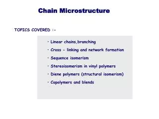
Types of Bone • Woven Bone • Lamellar Bone • Fibrolamellar Bone • Haversian Bone
Woven Bone- fast deposition • Found in fetal skeleton, areas of bone repair, or fast bone growth • Porous • Lamellar Bone- lower deposition rate, but more organized • Osteocytes reside in lamellar bone within the lacunae
Fibrolamellar Bone- speed and characteristics intermediate to woven and lamellar bone • Builds scaffolds quickly, then layer lamellar bone: alternating layer patterns • Haversian Bone- also known as Primary and Secondary Osteon • Central canal surrounded by series of lamellae • Canal carries arteries, nerves, and communicates with lacunae of concentric rings
Osteons • Bone tissue containing blood vessels surrounded by concentric rings of bone is called an osteon • Primary Osteon- formed by mineralization of cartilage, thus forming where bone is not present • Secondary Osteon- results from bone remodeling (osteoclastresorption, osteoblast deposition)
Primary Osteon Haversian Canal- carries vessels and nerves Lamellae- concentric rings made by collagen patterns Lacuna-osteocytes reside, turn over of bony matrix Canaliculi- link lacunae Cement- outer perimeter of an osteon Secondary Osteon Results from bone remodeling (osteoclastresorption, osteoblast deposition)
Bone Growth Cancellous Bone Synonymous for spongy bone Metaphyseal Remodeling Metaphyseal region decreases in diameter and incorporates into the long bone during growth - Produces distinct cortex stratification (layering)
Long Bone Growth Strategies Bone length limited by secondary center of ossification Can grow in length indefinitely
Stratified Cortex Produced by… • Metaphyseal reduction and reversal • Cortical drift • Tubercle formation • Secondary reconstruction (resorption and redeposition of bone) * Different parts of the same bone can have very different patterns
Cortical Stratification Example Indications: • Compacted coarse spongy bone: Tissue was produced during endosteal growth and was once located near the ends of the bone • Inner circumferential lamellae in bone cross section: Formed in the absence of spongy bone resulting from metaphyseal and diaphyseal remodeling
Femoral Growth in Length Mammalian Femur Cartilage formation Osteoblasts deposit bone over the cartilage surface Calcification of inner shaft, chondroclastresorb cartilage and vessels invade the cavity Osteoblast line cavity with bone Process continues along shaft • Bones thicken via periosteal bone production • Bones lengthen via terminal cartilage growth
Mammalian Femur Growth • 2 ossification centers: • Diaphysis • Spongy Bone • Contact of the two inhibits further growth
Cartilaginous regions usually degrade and don’t get preserved
Amprino’s Hypothesis • Italian scientist Amprino proposed that differences in bone tissue types reflect variations in bone depositional rates • Slow rate of bone formation results lamellar bone matrix- ordered arrangement of collagen fibers and osteocytes • Fast rate results in fibrolamellar bone tissue in which collagen fibers are haphazardly arranged and forms woven bone matrix. • Randomly distributed globular osteocytes and numerous entrapped blood vessels • Lamellar bone is eventually formed around each blood vessel to produce primary osteons
Rate of Bone Formation • Although organic components (collagen, osteocytes, and vessels) are not preserved, arrangement of crystals of the bone mineral is preserved • Bone mineral arrangement follows the organization of collagen matrix • Permits deduction about the type of bone tissue that was present • Inference about qualitative rate of bone formation • Fossil bones preserve canaliculi, lacunae, and cavities
Fibrolamellar bone Lamellar bone with lines of arrested growth Endosteallylamellated bone tissue
Quantitative information about the rate of bone formation: Fluorescent labeling using fluorochrome stains • Binds to newly formed bone • Used to determine where and how much bone was deposited • Different color stains to demarcate duration of differing periods of growth- quantification of bone deposition rate
Fluorochrome Staining Labeled in order: • Tetracyclin • Hematoporphyrine • DCAF TibialMetaphyseal Bone
Cyclical vs Noncyclical Growth • Structure of primary compact bone provides direct assessment of whether bone deposition was continuous or interrupted • Cyclic- interrupted growth • Non-cyclic- continuous growth
Cyclical Rates • Distinct growth rings or alternating bands of tissue types • This tissue type is termed zonal bone and the resulting bands of tissue are the zones and annuli • Zones more vascularized, represents fast growth • Poorly vascularizedannuli (usually lamellar bone) represents periods of slower growth • Pauses in bone growth are called Lines of Arrested Growth (LAG)
Cyclical vs Noncyclical Growth • Counting zones and annuli to estimate the organism’s age is called Skeletochronology Cyclic (interrupted) Noncyclic (continuous)
Bone Growth Rings • Reptiles generally have one single zone and annulus per year • Zones form during warmer months • Annulus form during unfavorable periods • Skeletochronology- age estimate • Dye patterns reveal seasonally related bone deposition • Spacing between rings become closer after sexual maturity • Estimates age of maturity
Zonal Bones Alligator Monitor Lizard Kangaroo Polar Bear
Growth Rings in Mammals • Unlike zonal bone formed in compact bone of reptiles, the “recording” part of the bone is the periosteal zone (outermost region of the coritcal bone) • Formed when overall body growth rate slowed down • Juvenile mammals have highly vascularized bone without any interruptions in the rate of growth • Later in ontogeny, mammals begin depositing dense bone periosteally and can form rest lines in this region • Only periosteal zone of compact bone is a recording structure of growth • Estimation of age requires correction factor
Zonal Bones Alligator Monitor Lizard Kangaroo Polar Bear
Complications • Mammal skeletochronology • Some mammals have periosteal bone without rest lines • Fast growing mammals • Periosteal bone is not considered good for age determination since it persists for a limited time only • Structure for recording growth is mainly cementum and dentine of teeth
Cementum is regarded as best for skeletochronology since it forms during entire of the animal and unaffected by remodeling and reconstruction • Dentine forms only until pulp caivty is filled • Affected by seasonal, intraseasonal, and daily changes in growth rate and calcification Cementum > Dentine
Noncyclical Bone Formation • Sustained, uninterrupted bone formation results in lack of zonation in the compacta • Indicates bone deposition was rapid and noncyclical • Tissue comprising the compacta can be entirely of fibrolamellar bone or can grade into a parallel-fibered or lamellar type of tissue • Fibrolamellar correlates with rapid growth
Bone Vascularization • Vascular bones have a network of blood vessels • Armand de Ricqles equated channels to be occupied fully by vascularization, which is wrong • Nerves and connective tissues also • Shape of cavity bore no resemblance to shape of blood vessels within • Avascularized bones have osteocytes developing a more extensive system of intercommunicating canaliculi (probably compensating for lack of blood vessels)
Types of Fibrolamellar Tissue • Laminar bone- bone formed in the periosteal region • Concentric channels around the cortex • Plexiform bone- rapid depositional bone with increased mechanical support • Concentric channel with radial connections • Reticular bone • Irregular channels
Blood Vessels • Simple Blood Vessels- vessels that have no change in bone microstructure around the vascular channel • Primary Osteon • Secondary Osteon • Primary osteons result when bone is formed rapidly and spaces are left around the blood vessels • These spaces are later filled by lamellar tissue around the blood vessel, thus can be distinguishable by a bony ring around each blood vessel
Secondary osteons do not reflect rate of bone formation, but are the result of secondary reconstruction • Removal of bone around a primary vascular canal, followed by subsequent redeposition of concentrically arranged lamellar bone in the erosion cavity • Secondary osteons are distinguished from primary by presence of a cement line or reversal line, marking the furthest extent of bone removal
Ontogenetic Status • Ontogeny describes the origin and development of an organism • Surface texture, articular surfaces, open/closed sutures • Sometimes, morphological features cannot distinguish ontogenetic status • Use of bone microstructure for more info • Juveniles typically have largely spongy bone from rapid rate of bone deposition • Result is woven bone with haphazardly arranged osteocytes
Woven bone provides more surface area for deposition of bone matrix than lamellar bone formation and permits more bone deposition at a given time • With increasing age, porous spaces are infilled with lamellar type of bone t issue and primary osteon results • Increases strength of developing bone
Sexing Bones • Hominids • Pelvic girdle and jaws exhibit sexual dimorphism • clueless about majority of extinct animals • Sinosauropteryx – preserved oviducts
Single taxa samples show dimorphism • Robust vs thin forms • Syntarsus • Perimedullary erosion cavities in robust forms linked to egg laying
Medullary bone • Found in the medullary cavity of ovulating birds • Mineral store for eggshell formation during egg laying Fig. 3.10 – femur of an ovulating hawk
Medullary bone might indicate female • Has not been recognized in any dinosaur bone • No evidence of medullary bone forming preovulation in modern reptiles Oviraptor found with a nest of eggs
Biomechanical Adaptations • Internal architecture of bone • Forces that may have acted on it during life • Muscle contraction during embryogenesis • Most important epigenetic factor of skeletal development • Bone is stressed and strained by complex factors • Muscular activity, joints, ligaments, and tendons
Mechanical response to forces • Dependent on • Magnitude force • Direction of force • Bone geometry • Bone material properties
Different bones respond differently • Variability in bone stiffness and strength • Type of bone tissue • Porosity • Mineralization • Location of bone in skeleton
Bone is a composite material • Mechanical properties of the whole bone is different from each of its components • Secondary osteons • Interstitial bone • Collagen-bone material
Mechanical properties are dependent on mineralization • High mineralization • Highly elastic but fractures easily • Low mineralization • Resistant to fractures but low elasticity
Mineralization is controlled by various factors • Mineralization lag time • Time delay between deposition of organic matrix and mineral • Newer bone is mechanically different than older bone • Bone remodeling • Removal of primary bone increases porosity
More factors • Metabolic bone diseases • Rickets • Lack of Vit D, Ca, Phosphates • Leads to soft weak bones • Osteomalacia • Lack of Vit D, cannot breakdown Vit D • leads to soft weak bones