Download
1 / 54
710 likes | 1.6k Vues
Skeletal Muscle (and a little cardiac) Excitation-Contraction Coupling. Ed Balog Applied Physiology 555 14 th St NW Rm 1303 ed.balog@ap.gatech.edu. [Ca 2+ ] 1-2 mM. Ca 2+. [Ca 2+ ] 100 nM. Na 2+. ADP + Pi. RyR. IP 3 R. Ca 2+. [Ca 2+ ] 1 mM. ATP. Endo/Sarcoplasmic Reticulum.

E N D
Skeletal Muscle (and a little cardiac) Excitation-Contraction Coupling Ed Balog Applied Physiology 555 14th St NW Rm 1303 ed.balog@ap.gatech.edu
[Ca2+] 1-2 mM Ca2+ [Ca2+] 100 nM Na2+ ADP + Pi RyR IP3R Ca2+ [Ca2+] 1 mM ATP Endo/Sarcoplasmic Reticulum ADP + Pi ATP Ca2+
Fertilization Stimulus-secretion coupling Metabolism Muscle Contraction Learning and Memory Gene regulation Cell death Immune cell activation Intracellular Calcium Signaling Berridge et al., Nature Rev. Mol. Cell Biol. 1:11, 2000.
Ca2+ Movement in Muscle Flux that increases cytoplasmic Ca2+ Flux that decreases cytoplasmic Ca2+ Cytoplasm Mitochondria Troponin & other Ca2+- binding proteins Ca2+ Buffers SR
1. Maximal Calcium Activated Force 2. Calcium Sensitivity 3. Calcium Delivered to Contractile Proteins Determinates of Contractile Force
Excitation-Contraction (EC) Coupling • The process linking depolarization of the muscle cell surface membrane to the release of Ca2+ from the sarcoplasmic reticulum (SR). • EC coupling controls the [Ca2+] within the muscle cell; [Ca2+] controls force.
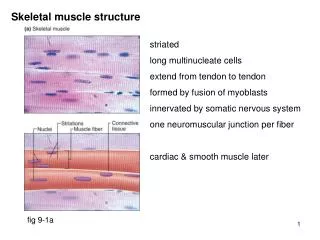
Sarcolemma Triad Transverse Tubule Sarcoplasmic Reticulum Skeletal Muscle Membrane System
Hypotheses: • Depolarization of the sarcoplasmic reticulum. • Chemical messenger from the transverse-tubules to the sarcoplasmic reticulum. • Ca2+-induced Ca2+ release (CICR). • Physical link between the transverse-tubules and the sarcoplasmic reticulum. This hypothesis also became to be known as depolarization-induced Ca2+ release (DICR)
Calcium-Induced Calcium Release? Small amount of Ca2+entering the cell from the extracellular fluid triggers much larger SR Ca2+release. Primary mechanism of EC coupling in the heart. Skeletal muscle SR Ca2+ release can be triggered via CICR under experimental conditions. But – Contraction can be elicited in the absence of extracellular Ca2+.
Physical Link? Primary mechanism in skeletal muscle. Electronmicrographs show electron-dense structures in triadic junction, called “feet” linking t-tubules and SR. Dysgenic mouse muscle lacking “feet” also lack EC coupling.
aka “Ca2+ release channel” or “junctional foot protein” Homotetramer; 2 million daltons. aka “DHPR”, voltage sensor” or “L-type channel” Heteromultimeric protein w/ 5 subunits. Origin of charge movement & L-type Ca2+ current. Excitation-contraction coupling: a tale of two Ca2+ channels Dihydropyridine Receptor DHPR tetrad T-tubule membrane RyR tetramer Ryanodine Receptor SR membrane
Lamb and Stephenson, Dept of Zoology, LaTrobe University High K+High Na+ Posterino, Proc Australian Physiol Pharmacol Soc 32: 28, 2001. Mechanically Peeled Muscle Fiber
Methods to Study Excitation-Contraction Coupling Fig. 5. Ca2+ sparks activated by membrane depolarization in skeletal muscle. The figure shows line scan images of sparks activated by small depolarizations (indicated at top) from a holding potential of −90 mV. For the pulses to −70 mV individual, randomly activated sparks are evident during the depolarization in the images and in the fluorescence records from individual, identified triads (below). The lowermost record in each column shows the average elevation of fluorescence from the entire image. The rightmost panel shows a depolarization to −60 mV during which the high frequency of Ca2+ sparks has resulted in a much larger elevation of fluorescence within the fiber, precluding the observation of individual sparks. Klein and Schneider Prog Biophys Mol Biol 92:308, 2006
Ryanodine [3H]Ryanodine Binding Ryanodine binds the open channel with high affinity and specificity. Binding reflects the open state of the channel.
Channels Lipid Bilayer Single Channel Recording in a Planar Lipid Bilayer
Some of the Whole-Cell Measurable Events of Excitation-Contraction Coupling
γ α2 C N N C N α1 δ S t-tubule lumen S C + + + + + + + + + + + + cytoplasm C N N β II-III loop and β-subunit vital for skeletal muscle ECC C DHPR (L-type Ca2+ Channel) • Member of voltage-gated ion channel superfamily, which also includes Na+ & K+ channels. • Pentamer; α1 subunit forms pore. • Cav1.1: skeletal muscle isoform; 1873 aa in humans • Cav1.2: cardiac isoform; 2169 aa in humans
10 μA/μF Cardiac 25 ms DHPR electrical signals:L-type calcium current • Cardiac current is larger and is fully activated by a ventricular action potential. Ca2+ influx via the cardiac DHPR activates RyR2 via Ca2+ induced Ca2+ release (CICR). • Skeletal muscle current is smaller and is barely activated during a skeletal muscle action potential. The current is not required for contraction. Skeletal Muscle
Skeletal Muscle Contraction Does Not Require the Entry of Extracellular Calcium Caputo & de Bolanos J Physiol 289: 175, 1979. Wang et al Biophys J 77:2709, 1999 2.5 mM Ca2+0 Ca2+ 2.5 mM Ca2+ Voltage dependence of Ica and contraction differ. Ica activation is too slow to contribute Ca entry during an action potential. Skeletal muscle can contract in the absence of extracellular Ca. Tetanus Twitch Dulhunty and Gage J Physiol 399:63, 1988
DHPR tetrad + + ΔV t-tubule membrane + + + + + DHPR electrical signals: Charge movement • Arises from movement of charged amino acids across membrane electric field. • Similar to ion channel gating currents, but larger and slower. • Required for skeletal muscle contraction.
Charge Movement & Ca2+ Release Top: voltage dependence of skeletal muscle contraction. A: Intracellular calcium transients recorded from a muscle fiber. B: T-tubule charge movement records from the same fiber. Below: Correlation between Charge movement and Ca2+ release rate. Caputo & de Bolanos J Physiol 289: 175, 1979.
Sarcolemma Triad Transverse Tubule Sarcoplasmic Reticulum Ryanodine Receptor (RyR) • The open channel binds Ryanodine, an alkaloid derived from the South American plant Ryania speciosa. • Member of intracellular Ca2+ channel family, includes IP3 receptor. • Largest known ion channel. • Three isoforms: RyR1 (skeletal), RyR2 (cardiac), RyR3 (wide cellular distribution, low abundance).
A I AL Calcium Dependence of RyR1 and RyR2 High affinity calcium binding site – Activates channel when bound. Calcium selective (KCa/KMg ~100) . A Low affinity calcium binding site – Inhibits channel when bound. Relatively unselective for divalent cations (KCa/KMg ~1) . I SR lumenal calcium binding site – Activates channel when bound. AL
RyR Macromolecular Complex Song et al Prog Biophys Mol Biol. 105:145, 2012.
RyR Channel Modulators (Partial List) Endogenous Adenine Nucleotides Calmodulin Mg2+ H+ Inorganic Phosphate Dihydropyridine Receptor FKBP12/12.6 Reactive Oxygen Species Nitric Oxide Exogenous Caffeine Ryanodine Ruthenium Red Volatile Anesthetics Depolarizing Muscle Relaxants Oxidizing/reducing agents Local Anesthetics 4-chloro-m-cresol Dantrolene
DHPRs α2 α1 δ γ T-tubule Membrane β Junctophilin RyR1 SR Membrane Arrangement of DHPR and RyR1 • 4 DHPRs per coupled RyR Serysheva et al PNAS 99:10370, 2002.
DHPR:RyR Arrangement and Ratio Varies with Muscle Fiber Type Toadfish Swim Bladder (very fast) DHPR tetrads Mammalian Fast-twitch Muscle RyR1 Mammalian Slow-twitch Muscle RyR2 Mammalian Cardiac Muscle Individual DHPRs
How are uncoupled skeletal muscle ryanodine receptor channels opened?
+ + + + + + + + + + + + + + + Ca2+-Induced Ca2+ Release Direct Coupling
Ca2+ DHPR tetrad (α1s) DHPR (α1c) + V t-tubule V + SR Ca2+ Ca2+ RyR2 RyR1 + + + + + + + Comparison of Cardiac and Skeletal Muscle Excitation-Contraction Coupling Skeletal Muscle: Mechanical Coupling Cardiac Muscle: Calcium-Induced Calcium Release Charge movement within DHPR and subsequent conformational change activates RyR via direct physical interaction. DHPR mediates Ca2+ influx, Ca2+ binds to and activates the underlying RyR.
Store-Operated Calcium Entry (SOCE) Requires depletion of the internal stores & has been best characterized in non-excitable cells. Requires STIM1 and ORAI Significant SR Ca2+ depletion required to reach activation threshold for SOCE only achieved during prolonged bouts of ECC. SOCE is not responsible for refilling the SR during periods of fiber quiescence. Excitation-Coupled Calcium Entry (ECCE) Activated following prolonged membrane depolarization Independent of the calcium stores. Requires functioning L-type channel and RYR1, but molecular identity of the pore remains undefined although it is likely to involve the L-channel. Two Forms of Ca2+ Entry in Skeletal Muscle
Store-Operated Ca2+ Channels Lewis Nature 446: 284, 2007.
Calcium Transporters in Muscle SERCA: Sarco/Endoplasmic Reticulum Calcium ATPase PMCA: Plasma Membrane Calcium ATPase NCX: Na/Ca Exchange MCU: Mitochondrial Uniporter
SERCA: Sarco/Endoplasmic Reticulum Calcium ATPase • Encoded by 3 mammalian genes • ~1000 amino acids • 10 TM helices • Located in intracellular organelles: ER and SR • 3 Cytoplasmic domains (A,N,P) • 2 Ca2+ transported per ATP hydrolyzed • Activity regulated by phospholamban and sarcolipin
SERCA Genes SERCA1 – Expressed in fast-twitch skeletal muscle. Two splice variants. SERCA1a in adult fast-twitch skeletal muscle. SERCA1b in embryonic skeletal muscle. SERCA2 – Expressed in cardiac and slow-twitch skeletal muscle. Three splice variants. SERCA2a in cardiac and slow-twitch skeletal muscle. SERCA2b low levels in most tissues (“house-keeper”) SERCA2c in embryonic heart cells and mesenchymal stem cells (give rise to muscle and bone) SERCA3 – Expressed in smooth muscle. Five splice variants. In smooth muscle, blood, and neural cells. Co-expressed with SERCA2b (and others). Why 3 genes with multiple splice variants? Tune enzyme activity (calcium affinity & maximal velocity) to cell type, ligand sites to modulate activity, binding sites for regulators, other unknown reasons.
PMCA:Plasma Membrane Ca2+-ATPase • Encoded by 4 mammalian genes • ~1300 amino acids • 10 TM helices • Located in surface and t-tubule membranes • 3 Cytoplasmic domains (A,N,P) • 1 Ca2+ transported out of cell per ATP hydrolyzed • Activity regulated by calmodulin Brini et al. FEBS J 280:5385, 2013
NCX:Sodium-Calcium Exchanger • Encoded by 3 mammalian genes • ~950 amino acids • 9 TM helices • Located in surface and t-tubule membranes • 1 Ca2+ transported out of cell per 3 Na+ into cell • Driven by Na+ potential (Em-ENa) • Reverses mode during depolarization Red = Calcium Green = Sodium
MCU:Mitochondrial Calcium Uniporter • A complex of proteins located in mitochondrial inner membrane. • Facilitated diffusion of Ca into matrix. • Driven by large electronegative potential (-180 mV). Marchi & Pinton, in press J Physiol 2013
RyR1 Malignant Hyperthermia Central Core Disease RyR2 Catecholaminergic Polymorphic Ventricular Tachycardia Arrhythmogenic Right Ventricular Dysplasia RyR3 None Identified, Yet Ryanodine Receptor Diseases
Malignant Hyperthermia An autosomal dominant pharmacogenetic disease characterized by an unusual metabolic reaction to volatile anesthetics and depolarizing muscle relaxants. Symptoms include: Hypercapnia Cyanosis Tachycardia Muscle rigidity Rhabdomyolysis Hyperthermia Incidence: 1 in 15,000 anaesthetized children 1 in 50,000 anaesthetized adults With the introduction of dantrolene the mortality rate has been reduced from ~80% to current ~5%. About 50% of cases linked to RyR1 mutations.
L13R C35R R 44C/H D60N Q155K R156K E160G R163C/L G165R D166N/G R177C Y178C G215E V218I M226K D227V G248R R316L R328W G341R R367L R401C/G/H I403M Q474H Y522S/C R530H R533C/H R533H R552W R614C/L S846L R1043C R1140C S1342G Q1589P P1592L S1728P/F M1729R P1787L M1814K A1832G G2060C V2117L D2129E R2163C R2163H/P V2168M A2200V T2206M/R V2210F V2212A V2214I V2280I I2321V R2336H N2342S E2344D V2346M E2348G A2350T R2355W F2364V P2366R A2367T G2375A A2428T D2431N/W G2434R R2435H/L A2437V R2452W/Q I2453T R2454C/H R2458C/H P2496L R2508G/C/H Y2510H E2545D V2550L R2591W T2596I R2676W D2730H/G G2733D T2787S R2840W E2880K E3104K R3119H R3350H K3367R P3527S E3583Q E3584Q R3707L Q3756E V3840I R3903Q I3916M D3986E G3990V S4050Y T4081M N4119Y Δ4124-4216 R4136S I4138T V4234L E4283V T4637A/I G4638D R4645Q Δ4647-4648 L4650P H4651P P4668S F4684S K4724Q Y4733E G4734E R4737W/G L4793P Y4796C F4808I L4814F I4817F G4820W L4824P R4825C/P T4826I L4838V V4849I A4856G F4860V R4861C/H Δ4863-4969 Y4864C K4876R M4880T G4891R R4893W/Q A4894T/V I4898T G4899R/E A4906V R4914G/T F4921S V4927F Δ4927-4928 I4938M D4939E A4940T G4942V F4960Y P4973L RyR1 MH/CCD Mutations 0 1000 2000 3000 4000 5000 Amino Acid Sequence
In Vitro Contracture Test Caffeine/Halothane Contracture Test European Malignant Hyperthermia Group (EMHG) Low threshold = contracture of ≥0.2 g at a concentration of ≤2 mM caffeine or ≤ 2% halothane MH Susceptible(MHS): Low contraction threshold for both Caffeine & Halothane MH Equivocal(MHE): Low threshold for one Normal (MHN): Normal threshold for both North American Malignant Hyperthermia Group (NAMHG) Low threshold = contracture of ≥0.3 g at a concentration of ≤2 mM caffeine or ≥0.3 g at ≤ 3% halothane MH Susceptible(MHS): Low contraction threshold for either Caffeine or Halothane Normal (MHN): Normal threshold for both Increased Sensitivity to RyR Activators Forms Basis for MH Screening R615C Homozygous R615C Heterozygous Normal Gallant and Lentz Am J Physiol 262:C422, 1992
How does abnormal Ca2+ regulation cause MH? MacLennan and Phillips Science 256:789, 1992.
Is there a link between MH and exertional heat illness? Porcine Stress Syndrome 26 soldier with exertional heat illness, all had positive in vitro contracture tests (Bendahan et al Anesth Anag 93: 683, 2001). 3 patients with positive IVC and RyR1 mutations; 2 had history of EHI (Brown et al Br J Anesth 88:508, 2002). Effectiveness of dantrolene in treatment of heat illness questioned (see Hadad et al Sports Med 34: 501, 2004). Y522S MH mutation knock-in mouse has an increased sensitivity to heat stress (Chelu et al FASEB J 20:329, 2006).
Cardiac Excitation Contraction Summary Bers 2002 Nature 425:198.