Pneumonia
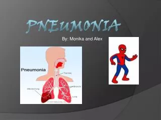
Pneumonia. Community acquired pneumonia . Definition. Pneumonia is acute infection leads to inflammation of the parenchyma of the lung ( the alveoli ) (consolidation and exudation ) The histologically Fibrinopurulent alveolar exudate seen in acute bacterial pneumonias.

Pneumonia
E N D
Presentation Transcript
Pneumonia Community acquired pneumonia
Definition • Pneumonia is acute infection leads to inflammation of the parenchyma of the lung (the alveoli) (consolidation and exudation) • The histologically • Fibrinopurulent alveolar exudate seen in acute bacterial pneumonias. • Mononuclear interstitial infiltrates in viral and other atypical pneumonias • Granulomas and cavitationseen in chronic pneumonias • It may present as acute, fulminant clinical disease or as chronic disease with a more protracted course
Epidemiology • Overall the rate of CAP 5.16 to 6.11 cases per 1000 persons per year • Mortality 23% • pneumonia are high especially in old people • Almost 1 million annual episodes of CAP in adults > 65 yrs in the US Risk factors • Age < 2 yrs, > 65 yrs • alcoholism • smoking • Asthma • prior influenza • HIV • Immuno suppression • institutionalization • Recent hotel : Legionella • Travel, pets, occupational exposures-birds(C-psittaci) • Aspiration • COPD • dementia
Etiological agents • Etiological agents of pneumonia could be bacterial, fungal, viral or parasitic or by other non-infectious factors like chemical, allergen
Pathogenesis Two factors involved in the formation of pneumonia • pathogens • host defenses.
Defense mechanism of respiratory tract • Filtration and deposition of environmental pathogens in the upper airways • Cough reflux • Mucociliary clearance • Alveolar macrophages • Humoral and cellular immunity • Oxidative metabolism of neutrophils
Pathophysiology : • Inhalation or aspiration of pulmonary pathogenic organisms into a lung segment or lobe. • Results from secondary bacteraemia from a distant source, such as Escherichia coli urinary tract infection and/or bacteraemia(less commonly). • Aspiration of Oropharyngeal contents (multiple pathogens).
Classification -Pathogen-(most useful-choose antimicrobial agents) -Anatomy -Acquired environment • Bacterial pneumonia • Streptococcus pneumoniae is the most frequently isolated pathogen • Typical • Gram-positive bacteria as • Streptococcus pneumoniae • Staphylococcus aureus • Group A hemolytic streptococci (2) Gram-negative bacteria - Klebsiellapneumoniae - Hemophilusinfluenzae - Moraxella catarrhal - Escherichia coli (3) Anaerobic bacteria
Viral pneumonia the most common cause of pneumonia in children < than 5 years - Adenoviruses - Respiratory syncytial virus • Influenza virus • Human metapneumovirus • SARS - Cytomegalovirus - Herpes simplex virus • Atypical pneumonia • Legionnaies pneumonia • Mycoplasma pneumonia • Chlamydophila pneumonia • Rickettsias • Francisellatularensis (tularemia), • Fungal pneumonia • Candida • Aspergilosis • Pneumocystis carnii Pneumonia caused by other pathogen • Parasites - protozoa
CAP and bioterrorism agents • Bacillus anthracis (anthrax) • Yersiniapestis (plague) • Francisellatularensis (tularemia) • C. burnetii (Q fever) • Level three agents
Classification by anatomy 1. Lobar: entire lobe 2. Lobular: (bronchopneumonia). 3. Interstitial
Classification by acquired environment • Community acquired pneumonia(CAP) • Hospital acquired pneumonia(HAP) • Nursing home acquired pneumonia (NHAP) • Immunocompromised host pneumonia (ICAP)
CAP-Cough/fever/sputum production + infiltrate • CAP : pneumonia acquired outside of hospitals or extended-care facilities • Streptococcus pneumoniae(most common) • Haemophilusinfluenzae • mycoplasmapneumoniae • Chlamydia pneumoniae • Moraxella catarrhalis • Drug resistance streptococcus pneumoniae(DRSP) is a major concern.
Classifications Typical Atypical Atypical’: not detectable on gram stain; won’t grow on standard media Mycoplasma pneumoniae Chlamydophilla pneumoniae Legionellapneumophila Influenza virus Adenovirus TB Fungi • Typical pneumonia usually is caused by bacteria • Strept. Pneumoniae • (lobar pneumonia) • S. aureus • Haemophilusinfluenzae • Gram-negative organisms • Moraxella catarrhalis
Community acquired pneumonia • Strep pneumonia 48% • Viral 23% • Atypical orgs(MP,LG,CP) 22% • Haemophilus influenza 7% • Moraxella catharralis 2% • Staph aureus 1.5% • Gram –ive orgs 1.4% • Anaerobes
Clinical manifestationlobar pneumonia • The onset is acute • Prior viral upper respiratory infection • Respiratory symptoms • Fever • shaking chills • cough with sputum production (rusty-sputum) • Chest pain- or pleurisy • Shortness of breath
Diagnosis • Clinical • History & physical • X-ray examination • Laboratory • CBC- leukocytosis • Sputum Gram stain- 15% • Blood culture-5-14% • Pleural effusion culture Pneumococcal pneumonia
Drug Resistant Strep Pneumoniae • 40% of U.S. Strep pneumoCAP has some antibiotic resistance: • PCN, cephalosporins, macrolides, tetracyclines, clindamycin, bactrim, quinolones • All MDR strains are sensitive to vancomycin or linezolid; most are sensitive to respiratory quinolones • β-lactam resistance - meningitis (CSF drug levels) • PCN is effective against pneumococcal Pneumonia at concentrations that would fail for meningitis or otitis media • For Pneumonia, pneumococcal resistance to β-lactams is relative and can usually be overcome by increasing β-lactam doses (not for meningitis!)
Pneumococcal CAP: Be cautious if using PCN if MIC >4. Avoid using PCN if MIC >8. • Remember that if MIC <1, pneumococcus is PCN-sensitive in sputum or blood (but need MIC <0.06 for PCN-sensitivity in CSF). MIC Interpretive Standards for S. pneumoniae. Clinical Laboratory Standards Institute (CLSI) 2011; 28:123.
Atypical pneumonia Approximately 15% of all CAP Not detectable on gram stain won’t grow on standard media Often extrapulmonary manifestations: Mycoplasma: otitis, nonexudativepharyngitis, watery diarrhea, erythemamultiforme, increased cold agglutinin titre Chlamydophilla: laryngitis Most don’t have a bacterial cell wall Don’t respond to β-lactams Therapy: macrolides, tetracyclines, quinolones (intracellular penetration, interfere with bacterial protein synthesis) • Chlamydia pneumonia • Mycoplasma pneumonia • Legionellaspp • Psittacosis (parrots) • Q fever (Coxiellaburnettii) • Viral (Influenza, Adenovirus) • AIDS • PCP • TB (M. intracellulare)
Mycoplasma pneumonia Mycoplasma pneumonia. Common people younger than 40. Crowded places like schools, homeless shelters, prisons. Usually mild and responds well to antibiotics. Can be very serious May be associated with a skin rash and hemolysis • Eaton agent (1944) • No cell wall • Mortality rate 1.4% • Rare in children and in > 65 • Myocarditis • Pancreatitis
Chlamydia pneumonia • Obligate intracellular organism • 50% of adults sero-positive • mild disease • Sub clinical infections common • 5-10% of community acquired pneumonia • Related to C psittacii • Budgies, parrots, pigeons and poultry • Birds often asymptomatic
Psittacosis • Chlamydophila psittaci • Exposure to birds • Bird owners, pet shop employees, vets • 1st: Tetracycline • Alt: Macrolide
Q fever • Coxiellaburnetti • Exposure to farm animals or parturient cats • 1st: Tetracycline, 2nd: Macrolide
Legionellapneumophila Legionnaire's disease. has caused serious outbreaks. Outbreaks have been linked to exposure to cooling towers ICU admissions. • Hyponatraemia common • (<130mMol) • Bradycardia • WBC < 15,000 • Abnormal LFTs • Raised CPK • Acute Renal failure • Urinary antigen
Symptoms Signs Minimal Few crackles Rhonchi Exhaustion Low grade fever • Insidious onset • Mild URTI to severe pneumonia • Headache • Malaise • Fever • dry cough • Arthralgia / myalgia
Diagnosis & Treatment • Macrolide • Rifampicicn • Quinolones • Tetracycline • Treat for 10-14 days • (21 in immunosupressed) • CBC • Mild elevation WBC • U&Es • Low serum Na (Legionalla) • Deranged LFTS • ↑ ALT • ↑AlkPhos • Cold agglutinins (Mycoplasma) • Serology • DNA detection
Differential diagnosis • Pulmonary tuberculosis • Lung cancer • Acute lung abecess • Pulmonary embolism • Noninfectious pulmonary infiltration
Evaluate the severity & degree of pneumonia Is the patient will require hospital admission? • Patient characteristics • Comorbid illness • Physical examinations • Basic laboratory findings
The diagnostic standard of sever pneumonia • Altered mental status • Pa02<60mmHg. PaO2/FiO2<300, needing MV • Respiratory rate>30/min • Blood pressure<90/60mmHg • Chest X-ray shows that bilateral infiltration, multilobar infiltration and the infiltrations enlarge more than 50% within 48h. • Renal function: U<20ml/h, and <80ml/4h
Patient Management • Outpatient, healthy patient with no exposure to antibiotics in the last 3 months • Outpatient, patient with comorbidity or exposure to antibiotics in the last 3 months • Inpatient : Not ICU • Inpatient : ICU
Antibiotic Treatment • Macrolide: Azithromycin, Clarithromycin • Doxycycline • Beta Lactam :Amoxicillin/clavulinic acid, Cefuroxime • Respiratory Flouroquinolone:Gatifloxacin, Levofloxacin or Moxifloxacin • Antipeudomonas Beta lactam: Cetazidime • Antipneumococcal Beta lactam :Cefotaxime
Antibiotic Treatment Treatment