Download
1 / 1
10 likes | 90 Vues
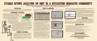
Explore dietary trends of a Byzantine monastic community through stable isotope analysis, offering intriguing insights into food consumption patterns and societal factors. Comparative studies reveal unique findings among global skeletal collections.

E N D
from St. Stephen’s adhered to an expected 15N path seen prior to weaning (Schurr 1998), followed by a rapid decline in the juveniles. For the St. Stephen’s remains, the 5 individuals less than 1 year of age showed elevated nitrogen levels, while the 6 subadults between 5-10 years old were significantly lower (p=0.04; df=9; t=1.7). Given the small sample size this is only a preliminary pattern, but worthy of further analysis. This study examined the dietary trends of a large monastic community in Jerusalem (5th-7th C AD). Previous studies of the Byzantine St. Stephen’s remains have shown a pattern of good nutrition, with little-to-no evidence of metabolic disorders, carious lesions, or calculus deposition (Sheridan 1999). Moreover, stature and robusticity for this largely male collection indicated a calorie-rich environment. The consumption of animal protein may account for such features, although historical texts for the time and region run counter to such an assessment. Thus, stable carbon and nitrogen isotope concentrations in bone were analyzed to help determine the composition of foods consumed by the monks of this wealthy urban community. Comparative Studies: The St. Stephen’s results were compared to numerous skeletal collections worldwide (Figure 5). Like the monks, the three Neolithic Greek sites of Alepotrypa, Tharrounia, and Theopetra shared a reliance on C3 plants (Papathanasiou 2003), however they diverged toward the lower end of the St. Stephen’s range for both 13C and 15N, indicating a stronger reliance on C3 plant consumption in the Greek communities. The Egyptian site of Ein Tirghi (ca. 800 BC to AD 350) showed a markedly different pattern from all other comparative collections, representing an arid region with unusually high nitrogen values (Dupras 1999). Although often associated with a diet dependent on marine resources, the nitrogen-rich soil in the surrounding area likely augmented the Ein Tirghi plant 15N values. Medieval Athens Ayios Nicolaos in Greece exhibited similarities to St. Stephen’s in their slightly elevated nitrogen levels, indicative of some animal protein consumption (Garvie-Lok 2001). Tell Leilan in Syria had a comparable 13C range to St. Stephen’s showing a reliance on C3 plants, but with lower nitrogen values. The archaeobotanical data suggested a heavy reliance on C3 plants, with little indication of meat consumption (Feasby 1998). The differences in 15N values between Tell Leilan and St. Stephen’s may indicate a higher animal protein component to the monastic diet. This project offers some intriguing patterns related to diet composition at Byzantine St. Stephen’s monastery. Although the male/female differences 15N are the likely result of physiological differences between the sexes, the strong subadult/adult divide is possibly related to weaning. Future work to enhance these data will include the isotopic analysis of archaeozoological samples from the site as well as an increased sample of subadults. Table 1.Distributions of ∂13C and ∂15N for the St. Stephen’s collection by sex and age. Stable Isotope Analysis: During photosynthesis, plants convert atmospheric CO2 into molecules containing either 3 or 4 carbon atoms. The more common C3 plants (trees and most vegetables) grow in temperate areas and have a 13C value of approximately -25 to -21‰. C4 varieties tend to be rarer and include maize, sorghum, and some grasses, with a 13C range of -15 to -5‰ (DeNiro 1985). Marine plants generally have an intermediate value and are reflected in the consumer animal (Figure 1). In addition, 15N values contribute to pinpointing specific aspects of dietary breadth. For instance, 15N concentrations are more positive in marine versus terrestrial organisms, since the food chain tends to be longer in seawater environments (Lambert 1997). Therefore, distinctly larger 15N values can suggest a predominantly marine-based diet. Consequently, when charted together, 13C and 15N values can identify an organism’s trophic level intake, such as terrestrial carnivores consum- ing C3 plant-eaters or marine carnivores feeding on vertebrates. Figure 2.Distributions of ∂13C and ∂15N values for the St. Stephen’s collection. Figure 5.Distributions of ∂13C and ∂15N values for several comparative collections. Monastic diet: According to historical records, the diet for most people during the Byzantine period was largely vegetarian. Bread composed a major portion of the daily intake; in fact, its production was of such high priority that Figure 1.Distributions of ∂13C and ∂15N in various trophic levels.Adapted from DeNiro (1985). The distribution of 13C and 15N values in relation to several comparative collections indicates a C3-based diet with some reliance on animal protein. This is corroborated by low incidence of carious lesions, minimal dental calculus deposits and attrition rates, overall stature and robusticity, a lack of skeletal markers of nutritional stress (in a region prone to complications such as growth retardation and porotic hyperostosis), and historical records of affluence at this large monastic complex. Textual sources detail a strict ascetical and vegetarian diet for the monks of Byzantine Palestine; evidence from the bones and teeth of the St. Stephen’s inhabitants suggest that the foods selected may have varied from this prescribed menu. granaries and ovens were located on most monastery grounds (Hirschfeld 1996). A variety of fruit and vegetables supplemented the diet of the monks, such as carobs, dates, figs, peas, lentils, and beans (Regnault 1999). Communities located in desert regions utilized wild products as well as garden domesticates, gathering an assortment of edible plants that enhanced dietary breadth. It is not known however, whether urban monasteries collected these particular plants (Hirschfeld 1992). Most of the recorded cereal grains consumed were of the C3 variety, as were the majority of fruit and vegetables. One possible exception was sorghum, a C4 grain that was commonly utilized by the poor for bread production (Hirschfeld 1995; Larsen 2000). Whether this is pertinent to the St. Stephen’s community remains unclear, as numerous historical documents remark on the affluence of what was the largest monastic complex in the region for nearly 100 years (Chitty 1966; Binns 1994). Meat held a unique status in both monastic and everyday Byzantine life. Most animal protein came from domesticated sheep, goats, and cattle as well as fish. Historical records emphasized stringent regulations accentuating the monks’ abstinence from consumption of this ‘luxury item.’ Some accounts concede that meat was given to “ailing monks,” while others claim that it was taboo under any circumstances (Hirschfeld 1992; Regnault 1999). Animal by-products such as cheese, eggs, and honey supposedly followed a similarly scarce pattern within monasteries, available only to the monks as gifts. Hirschfeld (1992) speculated that the scarcity of meat, rather than specific regulations against its consumption, may have accounted for its absence in monastic diets, particularly in the Judean desert. However, little information currently exists for urban monasteries. • Binns J. 1994. Ascetics and ambassadors of Christ. Oxford : Clarendon Press ; New York : Oxford University Press. • Chitty D. 1966. The desert a city. Crestwood, N.Y. : St. Vladimir's Seminary Press. • DeNiro M. 1985. Postmortem preservation and alteration of in vivo bone collagen isotope ratios in relation to paleodietary reconstruction. Nature 317:806‑809. • Dupras T. 1999. Dining in the Dakhleh Oasis, Egypt: Determination of diet using documents and stable isotope analysis. (Ph.D. Thesis, McMaster University, Canada). • Feasby R. 1998. Stable isotope evidence for dietary patterns and environmental conditions at Tell Leilan, Syria, ca. 1900-2900 B.C. (MA Thesis, University of Alberta, Canada). • Garvie-Lok S. 2002. Loaves and fishes: A stable isotope reconstruction of diet in medieval Greece. (Ph.D. Thesis, University of Calgary, Canada). • Hirschfeld Y. 1992. The Judean desert monasteries in the Byzantine period. New Haven and London: Yale University Press. • Hirschfeld Y. 1995. The Palestinian dwelling in the Roman-Byzantine period. Jerusalem: Israel Exploration Society. • Hirschfeld Y. 1996. The importance of bread in the deit of the monks of the Judean Desert. Byzantion, 66:143-155. • Keegan W. 1989. Stable isotope analysis of prehistoric diet. In: Iscan MY, Kennedy KAR, editors. Reconstruction of life from the skeleton. New York: Alan R. Liss, Inc. p 223-236. • Lambert J. 1997. Traces of the past: unraveling the secrets of archaeology through chemistry. Cambridge, MA: Perseus Publishing. • Papathanasiou A. 1999. A bioarchaeological analysis of health, subsistence, and funerary behavior in the eastern Mediterranean basin: A case study from Alepotrypa Cave, Greece. • Regnault L. 1999. The day-to-day life of the desert fathers in fourth-century Egypt. Petersham, Massachusetts: St. Bede’s Publications. • Schurr M. 1998. Using stable nitrogen-isotopes to study weaning behavior in past populations. World Archaeol 30:327-342. • Schurr M, Powell M. 2005. Role of changing childhood diets in the prehistoric evolution of food production: an isotopic assessment. Am J Phys Anthropol 126:278–294. • Sheridan S. 1999. New life the dead receive: The relationship between human remains and the cultural record. Rev Biblique 4:574-611. • Ubelaker D. 1999. Human skeletal remains: excavation, analysis, interpretation. 3rd edition. Washington, D.C.: Smithsonian Institution. Figure 3.Distributions of ∂13C and ∂15N values by sex. This investigation utilized 81 samples, including the left distal femur (n=53) and the left ilium (n=28). The total number of femora for which sex could be determined (n=38), based on bicondylar breadth and linea aspera robusticity, included 34 males and 4 females. For the ilia (n=12), sex determination using auricular surface elevation and sciatic notch width was possible for 8 males and 4 females. Subadult ilia (n=11) were divided into two age categories – infants (n=5) and juveniles (n=6) -- based on general size and lack of epiphyseal fusion (Ubelaker 1999). Sixty-nine individuals were classified as adults. Samples (0.1-0.2g) were manually and ultrasonically cleaned to remove dirt from the trabecular bone, and dried for forty-eight hours at 70ºC. They were soaked in 0.25 M hydrochloric acid for approximately two weeks and stirred daily. This demineralization stripped the bone of inorganic material. The remaining collagen was freeze-dried, then tested by elemental analyzer and stable isotope mass spectrometer to obtain 13C and 15N values. National Science Foundation Research Experiences for Undergraduates Institute for Scholarship in the Liberal Arts; University of Notre Dame L’École Biblique et Archéologique Française de Jérusalem Dennis Birdsell, Center for Environmental Science & Technology, University of Notre Dame Byzantine St. Stephen’s Project Laboratory for Biocultural Studies Department of Anthropology University of Notre Dame Melissa Regan1, Lesley Gregoricka1, Jaime Ullinger2, Mark Schurr1, and Susan Guise Sheridan1 1Department of Anthropology, University of Notre Dame 2Department of Anthropology, Ohio State University C/N ratios were within the acceptable 2.9-3.6 range for all 81 samples, indicating good protein preservation (Keegan, 1989). As seen in figure 2, the range of 13C was -20.6 to -15.4‰ (n=81; x=-19.0±0.7‰), and the 15N range was 7.2 to 14.7‰ (n=81; x=10.0±1.5‰). Table 1 includes all the 13C and 15N comparisons for the Byzantine St. Stephen’s collection. ∂13C ∂15N ∂N ∂N ∂C ∂C Variation by Sex: Statistical analysis of bicondylar breadth showed a highly significant difference between femora designated male and female (p=0.0005, df=36; t=3.5). The 13C range of all males fell between -20.6 and -15.4‰ (n=42; x=-19.0±0.8‰), -19.6 to -18.3‰ (n=8; x=-19.0±0.4) for the females. Male 15N values were 7.3 to 14.7‰ (n=42; x=9.9±1.4), females 7.5 to 11.6‰ (n=8; x=9.4±1.3‰). Figure 3 illustrates the male/female stable isotope distributions at St. Stephen’s. Male femora 13C ranged from -20.6 to -15.4‰ (n=34; x =-19.0±0.8), females from -19.6 to -18.3‰ (n=4; x=9.6±1.7‰). Male femur 15N fell between 7.3 and 14.7‰ (n=34; x =9.8±1.5‰), females from 7.5 to 11.6‰ (n=4; x=9.6±1.7‰). The male innominate 13C values were -20.0 to -18.5‰ (n=8; x=-19.1±0.5‰); females ranged from -19.3 to -18.5‰ (n=4; x=-19.0±0.3‰). Male and female innominate 15N values were 9.1 to 12.2‰ (n=8; x=10.6±1.1‰) and 8.1 to 10.7‰ (n=4; x=9.2±1.0‰), respectively. There was a significant difference in 15N values by sex for the innominates (p=0.02; df=10; t=2.2). These results might reflect a higher trophic level for male diets compared to females, but this is much more likely a sex-related metabolic difference (Schurr and Powell 2005).Also, it is important to note that sex determination for the ilia was limited to auricular surface elevation and sciatic notch width because all of the innominates were broken. The reliance on less specific sex determination methods is worth noting. The femora, assessed using both metric and non-metric sex indicators and a larger sample size, demonstrated no significant sex difference (p=0.4; df=36). Age Variation: Individuals were divided into three broad categories for comparison of stable isotope variation by age: infants, juveniles, and adults (Figure 4). The 13C ranged from -19.5 to -17.8‰ (n=5; x=-18.4±0.6‰) for infants, -19.6 to -18.4‰ (n=6; x= -18.9 ±0.4‰) for juveniles, and -20.6 to -15.4‰ (n=69; x=-19.0±0.7‰) for adults. The combined subadult (infants plus juveniles) 13C values spanned from -19.7 to -17.8 (n=12; x=-18.6 ±0.6‰). For 15N, infants ranged from 10.7 to 13.3‰ (n=5; x=12.6±1.0‰), juveniles from 7.2 to 11.3 (n=6; x=9.6±1.3‰), adults from 7.3 to 14.7‰ (n=69; x=9.8±1.3‰), and subadults from 7.2 to 13.3 (n=12; x=11.0±1.9‰). There was a significant difference by age for both stable isotopes in the St. Stephen’s collection. When combined as ‘subadults”, infants and juveniles showed a significant difference in 15N (p=0.03; df=79; t=1.7) and 13C (p=0.02; df=79;t=1.9) values compared to adults. The 15N differences between the two subadult groups resulted from the elevated infant nitrogen levels, reflecting a diet of breast milk enriched in 15N. The infant samples ∂N ∂C ∂N ∂C Figure 4.Distributions of ∂13C and ∂15N values by age (innominates only).