Download

1 / 27
270 likes | 297 Vues
Explore the intricate processes of nerve and muscle cell transmission, from dendrites to axons, action potentials, myelin sheaths, and muscle contractions. Learn the key concepts, types of fibers, and more.

E N D
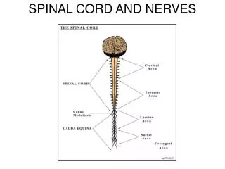
Cell body —> 5 - 7 dendrites —> Long, fibrous axon —> divides into terminal branches —> each ends in a terminal knob. Myelin sheaths - formed by Oligodendrocytes 4 zones to a nerve fiber Receptor (dendritic zone) A site where the propogated AP is generated An axonal process that transmits impulses to nerve endings The nerve endings which cause the release of synaptic transmitters The Nerve
Some Concepts • Resting Membrane Potential - primarily determined by 2 ions: Na+ and K+ • The membrane is much more permeable to K+ than Na+, therefore the resting membrane potential is closer to the equilibrium potential for K+ (- 70mV) • If a membrane potential becomes more positive than its resting membrane potential this is called depolarisation, the opposite is called hyperpolarisation • How is a resting membrane potential formed: https://www.khanacademy.org/science/biology/human-biology/neuron-nervous-system/a/the-membrane-potential
Action Potentials • Generation, transmission and propagation is a 7 step process • Step 1: Resting Membrane Potential • - 70mV (close to the K equilibrium potential) • Threshold of - 55mV is when the AP occurs (all or nothing) • Step 2: Threshold Potential • In response to a depolarising stimulus: some voltage gated Na Channels open and reach a threshold potential - at which point the K + Channels are overwhelmed
Step 3: Depolarisation • The entrance of Na causes a positive feedback loop and opens up more Na channels which generates the rapid upstroke. • Step 4: Equilibrium Potential • The membrane potential moves towards the equilibrium potential for Na + (+60 mV) but does not reach it during the action potential. • This increase in Na conductance is short lived, these channels rapidly enter a closed state called the inactivated state and remain in this state for a few milliseconds before they can be reactivated (ABSOLUTE REFRACTORY PERIOD)
Step 5: Repolarisation • Because of the overshoot the membrane potentials are reversed and so the flow of Na is also reversed. • Voltage gated K channels open. • These two factors contribute to repolarisation • The opening of voltage gated K channels is slower and more prolonged than the opening of the Na + channels and consequently, much of the increase in K conductance comes after the Na conductance. • Step 6: Hyperpolarisation • The net movement of K out of the cell helps complete the process of repolarisation. • Step 7: Return to Resting Membrane Potential.
Some Concepts • Refractory Period • Absolute: from firing until 1/3rd of the way through repolarisation no amount of stimulation will cause an AP to be triggered • Relative: a stronger than normal stimulus may trigger an AP • All or nothing • The minimal intensity of stimulating current will elevate the resting membrane potential to a threshold potential • Once threshold potential is reached further increases in this stimulating potential produce no further increment increases in the AP • If the stimulating threshold is sub-threshold, there is no AP generated • Electrogenesis of the AP • The affected cell’s polarity is reversed, so positive charge is on the inside and -ve charge on the outside. The adjacent normal membrane’s positive charge flows into this current sink. This causes depolarisation of the adjacent cells.
Some Concepts • Saltatory (“to hop/leap”) Conduction: The movement of current from one Node of Ranvier to the next. Occurs due to the myelinated segments between Nodes of Ranvier. • Allows conduction to move 50 times faster.
Muscles • 3 groups of muscle • Skeletal • Large mass • Cross striations and contains T tubules • Does not contract in absence of nervous stimulation, under voluntary control • Type 1 - slow, red; high oxidative capacityType 2 - fast/white/glycolytic, fast ATPase rate low oxidative capacity • Cardiac • Cross striations • Functionally syncytial • Contracts rhythmically in the absence of external innervation due to the presence of pacemaker cells • Smooth • Lacks cross striations • Found in hollow viscera • Functionally syncyitical and contains pace makers that discharge irregularly • Can remain in continuously contracted ie. “latched” state without energy requirement
Skeletal Muscle • Shortening of contractile elements in muscle is brought about by a sliding of thin filaments over thick filaments. • Depolarisation is due to Na influx and repolarisation is due to K efflux which occurs from a release of Ach at the motor end plate which binds to nicotinic receptorson the post synaptic junction. • T tubules propagate the action potential into the muscle fibers. • SacroplasmicReticulum releasesCa 2+ • Calcium binds troponin C, uncovering the myosin binding site on the actin. • Actin and myosin bind, thick and thin filaments move-there is a power stroke. • ATP binds and the actin/myosin detach and as ATP turns to ADP the myosin returns to its original position. • Ca is pumped back into the cell using an ATP driven mechanism.
Cardiac Muscle • Muscle fibers branch and interdigitate but is each complete unit • One nucleus • The cell borders are held by intercalated discs - these provide a strong union between fibres, and good force transmission • Along the side of fibres there are GAP JUNCTIONS which provide low resistance bridges for the spread of excitation to contract as though they were a SYNCYTIUM
Cardiac Muscle • Correlation between length and strength • In the heartthe degree of stretch is determined by the diastolic filling. • The pressure developed in the ventricle is proportional to the total tension developed. This is due to the fibers moving apart creating active tension. This is STARLING’S LAW. • The developed tension increases as volume increases up to a certain point and then begins to decrease due to a disruption of cardiac muscle fibers. • Force contraction is also increased by catecholamines mediated by beta 1 receptors and cAMP —> essentially leads to phosphorylation of Ca channels allowing them to be open for a greater period of time.
Cardiac Muscle • Metabolism - largely reliant of aerobic metabolism • Under basal conditions • 35% carbs • 5% ketones and amino acids • 60% fat
Cardiac Pacemaker • Characterised by absence of Na channels so that membrane potentials slowly, rather than rapidly, rise as voltage gated Ca channels are activated • At the peak of depolarisation K begins to flow out causing depolarisation. The K current begins to slow as the cell becomes hyper polarised • This triggers the funny (H) channel which is a Na/K channel and this starts the pre potential • The Ca channels then open, at first a transient (T) channel completes the pre potential and then an L channel opens causing the depolarisation
Pacemaker AP • Characterised by automaticity • SA node • AV node • Bundle of HiS
Nervous system and the cardiac action potential • Parasympathetic - increased Ach, opens K channels and slows opening of Ca channels causing hyperpolarisationof the membrane and a decrease in the AP slope. • Sympathetic - via increased noradrenaline (acting onbeta receptors)increases opening of funny (H) channels and L type Ca channels therefore speeds up both the depolarisation of the pre-potential and increases the firing rate and rateof the depolarisation impulse too.
Smooth Muscle • No striations • Glycolysis mainly for energy • Spontaneous activity in absence of nervous stimulation • Initiation of contraction is due to Ca influx • Unstable membrane potential causing continuous irregular contraction independent of nerve supply • In visceral smooth muscle stretch triggers depolarisation and contraction
Ca2+ binds calmodulin which activates calmodulin dependent myosin light chain kinase which phosphorylates myosin light chains. This allows for myosin crosslinking bridges to form with actin.