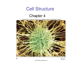
Cell Structure
Cell Structure. Chapter 3. What is a cell?. Cells are the smallest unit of life. Though there are many different kinds of cells, all cells share certain features, including: A plasma membrane Cytoplasm (with ribosomes) DNA. Plasma Membrane.

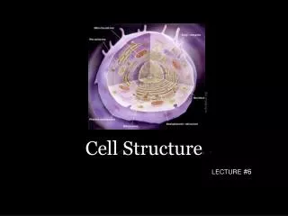
Cell Structure
E N D
Presentation Transcript
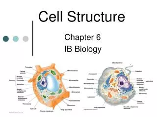
Cell Structure Chapter 3
What is a cell? • Cells are the smallest unit of life. • Though there are many different kinds of cells, all cells share certain features, including: • A plasma membrane • Cytoplasm (with ribosomes) • DNA
Plasma Membrane • Separates the activities taking place inside of the cell from the outside of the cell • Composed of a lipid bilayer (two layers of phospholipids) • Is selectively permeable (allowing some things through but not others)
Cytoplasm • Fluid or jellylike mixture contained within the cell • Most of the cell’s metabolic activities take place in the cytoplasm • Contains organelles in many cells (but not all)
DNA • Contains the cells hereditary information • All cells begin life with DNA but some lose it as they mature (ie., red blood cells) • Prokaryotes- DNA is suspended and concentrated in a region in cell cytoplasm called the nucleoid • Eukaryotes- DNA is contained within a double membrane bound structure called the nucleus
Almost all cells are too small to see with the naked eye. Why? • Cell size is limited by its surface-to-volume ratio. • Cells must exchange materials with their environment (nutrients enter the cell and wastes exit across the plasma membrane). • Cells must be able to permit this exchange fast enough to supply molecule -building activities with necessary nutrients and excrete wastes fast enough to prevent the cell from being poisoned. • Since a cells volume increases faster than its plasma membrane’s surface area as its size increases, the plasma membrane of a cell whose size increased very much would not be able to permit the exchange of nutrients and wastes fast enough to keep the cell alive
Surface-to-volume Ratio A B C Surface area = # of sides x (length of side)2 Volume = (length of side)3 What is the surface area to volume ratio for Cell A? Cell B? Cell C?
The Cell Theory • Every living organism is made of one or more cells. • A cell is the smallest unit of life, individually alive even as part of a multi-celled organism. • Every living cells arises from the division of pre-existing cells. • Cells contain hereditary material (DNA) that they pass on to their offspring during cell division.
Discovering Cells • Most cells are so small that they cannot be observed with the naked eye. Therefore, cells are generally measured in micrometers (10-6 m or one –thousandth of a millimeter or one-millionth of a meter). • Because cells are so small, they were not discovered until the microscope was invented. • Anton van Leeuwenhoek invented the first microscope. • He called the bacteria that he saw as he viewed tartar scrapings from his teeth “animalcules” and “beasties”. • Robert Hooke modified Leeuwenhoek’s microscope by adding another lens, making it easier to use to visualize smaller objects. Modern microscopes are still based on his design.
Discovering cells • Hooke, viewing thin slices of cork, called the compartments he saw “cellulae” and thus, ther term “cell” was coined. • In the mid 1800’s, scientists Schleiden and Schwann, realized that cells are independent living units, even when they are part of the tissues of animals or plants. • These two scientists states the cell theory in 1839.
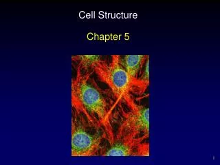
Modern Microscopes • Some modern microscopes use light to help us visualize objects (as did Leeuwenhoek’s and Hooke’s microscopes). Generally, specimen must be stained so that more detail is visible when a light microscope is used. • A fluorescence microscope involves researchers attaching a light-emitting tracer to a molecule or cell so that it will fluoresce when a laser beam from the microscope is focused on it.
Modern Microscopes Image from a fluorescent microscope Image from a light microscope
Modern Microscopes • Electron microscopes can reveal even finer details. These microscopes use a beam electrons (rather than light) to illuminate a specimen. • A transmission electron microscope (TEM) beam electrons through a thin specimen, revealing internal details as shadows on the resulting image • A scanning electron microscope (SEM) directs a beam of electrons back and forth across the surface of a specimen that has been coated with a thin layer of a metal (such as gold). This metal gives off electrons and x rays, forming an image of the surface of the specimen. • Both kinds of electron microscopes give us much more resolution that do light microscopes, allowing us to visualize objects as small as 2 nanometers.
Modern Microscopes • One drawback of using electron microscopes is that they produce black and white images only. • Since light is not being used, there is no color. • Another drawback is that only dead specimens may be viewed with an electron microscope. • Since water would interfere with the beam of electrons used by electron microscopes, any specimen being viewed with an electron microscope cannot contain water. Therefore, it must be dead.
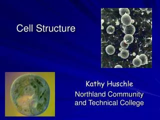
Modern Microscopes Image from an SEM Image from a TEM
The Structure of the Plasma Membrane • A double layer of phospholipids. • Phospholipids contain a polar hydrophillic (water loving) phosphate “head” with two nonpolar hydrophobic (water fearing) lipid “tails” attached.
The Structure of the Plasma Membrane • The nonpolar lipid tails associate with one another while the polar phosphate tails associate with the water on the inside and outside of the cell, forming the cell membrane. • The lipid bilayer also has other molecules, such as steriods (cholesterol) and proteins, embedded. • Cholesterol contributes to the stability of the cell membrane. More cholesterol causes the membrane to be more stiff. • Proteins allow the membrane to be selectively permeable by forming pores and channels through which certain molecules can enter or leave the cell. These proteins also enable the cell to control some materials that enter or leave the cell. • These molecules float around within the plasma membrane. The membrane is said to be fluid. This is called the fluid mosaic model of the plasma membrane.
Plasma Membrane Proteins • Enzymes: proteins in the plasma membrane that accelerate chemical reactions without themselves being changed • Adhesion proteins: attach cells to each other in animal tissues • Recognition proteins: identity “tags” that enable your body’s immune system to recognize your cells as your own, allowing it to also recognize cells that are not your own so that they can be attacked and destroyed. • Receptor proteins: bind to special chemical signals outside the cell (such as a hormone), which triggers a change in the cell’s activities, causing the cell to do something that it hadn’t previously done before. These changes can involve metabolism, movement, division, or even cell death. • Transport proteins: move specific substances across the cell membrane, usually by means of a channel.
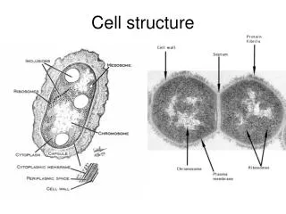
Prokaryotic cells = Bacteria • “Prokaryote” = “before the nucleus” • All are single-celled. • Are the smallest and most diverse cells. • Inhabit nearly every environment on Earth • Make up the Bacteria and Archae domains of the three domain classification system • DNA is contained in a single, circular chromosome called a nucleoid, usually not enclosed by a cell membrane • May contain small circles of DNA called plasmids, which carry a few genes, such as those that can confer antibiotic resistance
Prokaryotic cells • Many contain one or more flagella, structures that extend from the plasma membrane that rotate like a propeller, allowing the cell to move • Many also contain pili extending from their surface. These structures help the cell to cling to a surface, such as a another bacterial cell. The cell can then transfer a plasmid through the pilus to the other bacterial cell to which it is attached. • All also contain a cell wall made of peptides (proteins) and polysaccharides (sugars) surrounding the plasma membrane. Polysaccharides make the cell sticky so that it can stick to surfaces. It also offers some protection against predators and toxins.
Prokaryotic cells • The plasma membrane of prokaryotes contains transporter and receptor proteins that make it selectively permeable, just like eukaryotic cells. • It also contains proteins that are important for metabolic processes, such as the proteins that are necessary for carrying out photosynthesis found in the plasma membrane of cyanobacteria.