Download
1 / 29
300 likes | 516 Vues
Marieb Chapter 13 Part C. Innervation of Skeletal Muscle. Takes place at a neuromuscular junction Neurotransmitter ? NT binds to receptors, resulting in: Movement of Na + and K + across the membrane Depolarization of the muscle cell An graded potential, which triggers an action potential.

E N D
Innervation of Skeletal Muscle • Takes place at a neuromuscular junction • Neurotransmitter ? • NT binds to receptors, resulting in: • Movement of Na+ and K+ across the membrane • Depolarization of the muscle cell • An graded potential, which triggers an action potential
Myelinated axon of motor neuron Action potential (AP) Axon terminal of neuromuscular junction Nucleus 1 Action potential arrives at axon terminal of motor neuron. Sarcolemma of the muscle fiber 2 Voltage-gated Ca2+ channels open and Ca2+ enters the axon terminal. Ca2+ Synaptic vesicle containing ACh Ca2+ 3 Ca2+ entry causes some synaptic vesicles to release their contents (acetylcholine) by exocytosis. Mitochondrion Axon terminal of motor neuron Synaptic cleft Fusing synaptic vesicles Junctional folds of sarcolemma 4 Acetylcholine, a neurotransmitter, diffuses across the synaptic cleft and binds to receptors in the sarcolemma. ACh Sarcoplasm of muscle fiber K+ Na+ Postsynaptic membrane ion channel opens; ions pass. 5 ACh binding opens ion channels that allow simultaneous passage of Na+ into the muscle fiber and K+ out of the muscle fiber. Degraded ACh ACh Postsynaptic membrane ion channel closed; ions cannot pass. Na+ 6 ACh effects are terminated by its enzymatic breakdown in the synaptic cleft by acetylcholinesterase. K+ Acetylcholinesterase Figure 9.8
Innervation of Visceral Muscle and Glands • Autonomic motor endings and visceral effectors are simpler than somatic junctions • Branches form synapses via varicosities • Acetylcholine and norepinephrine act indirectly via second messengers • Visceral motor responses are slower than somatic responses
Varicosities Autonomic nerve fibers innervate most smooth muscle fibers. Smooth muscle cell Varicosities release their neurotransmitters into a wide synaptic cleft (a diffuse junction). Synaptic vesicles Mitochondrion Figure 9.27
Reflexes • Inborn (intrinsic) reflex: a rapid, involuntary, predictable motor response to a stimulus • Learned (acquired) reflexes result from practice or repetition, • Example:
Reflex Arc • Components of a reflex arc (neural path) • —site of stimulus action • —transmits afferent impulses to the CNS • —either monosynaptic or polysynaptic region within the CNS • —conducts efferent impulses from the integration center to an effector organ • —muscle fiber or gland cell that responds to the efferent impulses by contracting or secreting What is this an example of?
Stimulus Skin Interneuron 1 Receptor 2 Sensory neuron 3 Integration center 4 Motor neuron 5 Effector Spinal cord (in cross section) Figure 13.14
Spinal Reflexes • Spinal somatic reflexes • Integration center is in the spinal cord • Effectors are skeletal muscle • Why do they exist?
Stretch and Golgi Tendon Reflexes • For skeletal muscle activity to be smoothly coordinated, proprioceptor input is necessary • tell muscle length • tell muscle and tendon tension
Secondary sensory endings Efferent (motor) fiber to muscle spindle Primary sensory endings Muscle spindle Intrafusal muscle fibers Sensory fiber Golgi tendon organ Tendon Figure 13.15
Muscle Spindles • Excited by stretch of muscle and muscle spindle • Stretch causes an increased rate of impulses in sensory fibers • Motor fibers then cause muscle contraction • What kind of feedback?
Stretch Reflexes • Maintain muscle tone in large postural muscles • Cause muscle contraction in response to increased muscle length (stretch)
Stretch Reflexes • How a stretch reflex works: • Stretch activates the muscle spindle • Sensory neurons synapse directly with motor neurons in the spinal cord • Motor neurons cause the stretched muscle to contract • All stretch reflexes are monosynaptic and ipsilateral
Stretch Reflexes • Reciprocal inhibition also occurs—Sensory fibers synapse with interneurons that inhibit the motor neurons of antagonistic muscles • Example: In the patellar reflex, the stretched muscle (quadriceps) contracts and the antagonists (hamstrings) relax • Can you think of another example?
Stretched muscle spindles initiate a stretch reflex,causing contraction of the stretched muscle andinhibition of its antagonist. The events by which muscle stretch is damped The sensory neurons synapse directly with motor neurons (red), which excite the stretched muscle. Afferent fibers alsosynapse with interneurons (green) that inhibit motorneurons (purple) controlling antagonistic muscles. 2 When muscle spindles are activatedby stretch, the associated sensoryneurons (blue) transmit afferent impulsesat higher frequency to the spinal cord. 1 Sensoryneuron Cell body ofsensory neuron Initial stimulus(muscle stretch) Spinal cord Muscle spindle Antagonist muscle Figure 13.17 (1 of 2), step 2
The patellar (knee-jerk) reflex—a specific example of a stretch reflex 2 Quadriceps(extensors) 3a 3b 3b 1 Patella Musclespindle Spinal cord(L2–L4) Tapping the patellar ligament excitesmuscle spindles in the quadriceps. 1 Hamstrings(flexors) Patellarligament 2 Afferent impulses (blue) travel to thespinal cord, where synapses occur withmotor neurons and interneurons. 3a The motor neurons (red) sendactivating impulses to the quadricepscausing it to contract, extending theknee. +– Excitatory synapseInhibitory synapse 3b The interneurons (green) makeinhibitory synapses with ventral horn neurons (purple) that prevent theantagonist muscles (hamstrings) fromresisting the contraction of thequadriceps. Figure 13.17 (2 of 2)
Golgi Tendon Reflexes • Polysynaptic reflexes • Help to prevent damage due to excessive stretch • Important for smooth onset and termination of muscle contraction
Golgi Tendon Reflexes • Too much contraction? • Contraction activates Golgi tendon organs • Afferent impulses are transmitted to spinal cord • Contracting muscle relaxes and the antagonist contracts (reciprocal activation) • Information transmitted simultaneously to the cerebellum is used to adjust muscle tension
2 1 Afferent fibers synapse with interneurons in the spinal cord. Quadriceps strongly contracts. Golgi tendon organs are activated. Interneurons Quadriceps (extensors) Spinal cord Golgi tendon organ Hamstrings (flexors) 3b 3a Efferent impulses to antagonist muscle cause it to contract. Efferent impulses to muscle with stretched tendon are damped. Muscle relaxes, reducing tension. + Excitatory synapse – Inhibitory synapse Figure 13.18
Flexor and Crossed-Extensor Reflexes • Flexor (withdrawal) reflex • Initiated by a painful stimulus • Causes automatic withdrawal of the threatened body part • Ipsilateral and polysynaptic
Flexor and Crossed-Extensor Reflexes • Crossed extensor reflex • Occurs with flexor reflexes in weight-bearing limbs to maintain balance • Consists of an ipsilateral flexor reflex and a contralateral extensor reflex • The stimulated side is withdrawn (flexed) • The contralateral side is extended
Let’s Combine The Two! + Excitatory synapse – Inhibitory synapse Interneurons Efferent fibers Afferent fiber Efferent fibers Extensor inhibited Flexor inhibited Arm movements Flexor stimulated Extensor stimulated Site of reciprocal activation:At the same time, the extensor muscles on the opposite side are activated. Site of stimulus: a noxious stimulus causes a flexor reflex on the same side, withdrawing that limb. Figure 13.19
Superficial Reflexes • Elicited by gentle cutaneous stimulation • Depend on central or spinal reflex arcs
Superficial Reflexes • Plantar reflex • Stimulus: stroking lateral aspect of the sole of the foot • Response: downward flexion of the toes • Tests for function of corticospinal tracts
Superficial Reflexes • Babinski’s sign • Stimulus: same as in previous slide • Response: dorsiflexion of big toe and fanning of toes • Present in infants due to incomplete myelination • In adults, indicates corticospinal or motor cortex damage
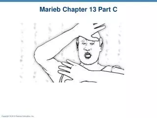
Plantar vs. Babinski Reflex Normal or Abnormal?
Superficial Reflexes • Abdominal reflexes • Cause contraction of abdominal muscles and movement of the umbilicus in response to stroking of the skin • Vary in intensity from one person to another • Absent when corticospinal tract lesions are present