Marieb Chapter 22: The Respiratory System Part A
390 likes | 1.25k Vues
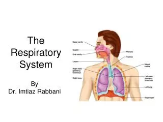
Marieb Chapter 22: The Respiratory System Part A. Respiration. Pulmonary ventilation (breathing): movement of air into and out of the lungs External respiration: O 2 and CO 2 exchange between the lungs and the blood Transport: O 2 and CO 2 in the blood

Marieb Chapter 22: The Respiratory System Part A
E N D
Presentation Transcript
Respiration • Pulmonary ventilation (breathing):movement of air into and outof the lungs • External respiration: O2 and CO2exchange between the lungsand the blood • Transport: O2 and CO2in the blood • Internal respiration: O2 and CO2exchange between systemic bloodvessels and tissues Respiratory system Circulatory system
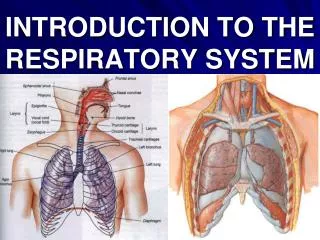
Functional Anatomy • Respiratory zone: site of gas exchange • Microscopic structures: respiratory bronchioles, alveolar ducts, and alveoli • Conducting zone: passageways to gas exchange sites • Includes all other respiratory structures • Respiratory muscles: diaphragm and other muscles that promote ventilation
Bronchi and Subdivisions • Air passages undergo 23 orders of branching • Branching pattern called the bronchial (respiratory) tree
Respiratory Zone • Respiratory bronchioles, alveolar ducts, alveolar sacs (clusters of alveoli) • ~300 million alveoli account for most of the lungs’ volume and are the main site for gas exchange
Alveoli Alveolar duct Respiratory bronchioles Alveolar duct Terminal bronchiole Alveolar sac (a) Figure 22.8a
Respiratory Membrane • ~0.5-m-thick air-blood barrier • Alveolar and capillary walls and their fused basement membranes • Alveolar walls • Single layer of squamous epithelium (type I cells) • Scattered type II cuboidal cells secrete surfactant and antimicrobial proteins
Red blood cell Nucleus of type I (squamous epithelial) cell Alveolar pores Capillary O2 Capillary Type I cell of alveolar wall CO2 Alveolus Macrophage Alveolus Endothelial cell nucleus Alveolar epithelium Fused basement membranes of the alveolar epithelium and the capillary endothelium Respiratory membrane Red blood cell in capillary Alveoli (gas-filled air spaces) Type II (surfactant- secreting) cell Capillary endothelium (c) Detailed anatomy of the respiratory membrane Figure 22.9c
Terminal bronchiole Respiratory bronchiole Smooth muscle Elastic fibers Alveolus Capillaries (a) Diagrammatic view of capillary-alveoli relationships Figure 22.9a
Alveoli • Surrounded by fine elastic fibers • Contain open pores that • Connect adjacent alveoli • Allow air pressure throughout the lung to be equalized • House alveolar macrophages that keep alveolar surfaces sterile
Fluids And The Lung • Pleural fluid fills the pleural cavity • Provides lubrication and surface tension • Surfactant within the alveoli • Lowers alveolar surface tension
Mechanics of Breathing • Pulmonary ventilation consists of two phases • Inspiration: gases flow into the lungs • Expiration: gases exit the lungs
Pressure Relationships in the Thoracic Cavity • Atmospheric pressure (Patm) • Pressure exerted by the air surrounding the body • 760 mm Hg at sea level • Respiratory pressures are described relative to Patm • Negative respiratory pressure is less than Patm • Positive respiratory pressure is greater than Patm • Zero respiratory pressure = Patm
Intrapulmonary Pressure • Intrapulmonary (intra-alveolar) pressure (Ppul) • Pressure in the alveoli • Fluctuates with breathing • Always eventually equalizes with Patm
Intrapleural Pressure • Intrapleural pressure (Pip): • Pressure in the pleural cavity • Fluctuates with breathing • Always a negative pressure (<Patm and <Ppul) • What happens if it equalizes with atmospheric pressure?
Intrapleural Pressure • Negative Pip is caused by opposing forces • Two inward forces promote lung collapse • Elastic recoil of lungs decreases lung size • Surface tension of alveolar fluid reduces alveolar size • One outward force tends to enlarge the lungs • Elasticity of the chest wall pulls the thorax outward
Alveolar Surface Tension • Surfactant • Detergent-like lipid and protein complex produced by type II alveolar cells • Reduces surface tension of alveolar fluid and discourages alveolar collapse • Insufficient quantity in premature infants causes infant respiratory distress syndrome
Atmospheric pressure Parietal pleura Thoracic wall Visceral pleura Pleural cavity Transpulmonary pressure 760 mm Hg –756 mm Hg = 4 mm Hg 756 Intrapleural pressure 756 mm Hg (–4 mm Hg) 760 Intrapulmonary pressure 760 mm Hg (0 mm Hg) Lung Diaphragm Figure 22.12
Homeostatic Imbalance • Atelectasis (lung collapse) is due to • Plugged bronchioles collapse of alveoli • Wound that admits air into pleural cavity (pneumothorax)
Pulmonary Ventilation • Inspiration and expiration • Mechanical processes that depend on volume changes in the thoracic cavity • Volume changes pressure changes • Pressure changes gases flow to equalize pressure
Boyle’s Law • The relationship between the pressure and volume of a gas • Pressure (P) varies inversely with volume (V): P1V1 = P2V2
Inspiration • An active process • Inspiratory muscles contract • Thoracic volume increases • Lungs are stretched and intrapulmonary volume increases • Intrapulmonary pressure drops (to 1 mm Hg) • Air flows into the lungs, down its pressure gradient, until Ppul = Patm
Changes in lateral dimensions (superior view) Changes in anterior- posterior and superior- inferior dimensions Sequence of events 1 Inspiratory muscles contract (diaphragm descends; rib cage rises). Ribs are elevated and sternum flares as external intercostals contract. Thoracic cavity volume increases. 2 External intercostals contract. 3 Lungs are stretched; intrapulmonary volume increases. Intrapulmonary pressure drops (to –1 mm Hg). 4 5 Air (gases) flows into lungs down its pressure gradient until intrapulmonary pressure is 0 (equal to atmospheric pressure). Diaphragm moves inferiorly during contraction. Figure 22.13 (1 of 2)
Expiration • Quiet expiration is normally a passive process • Inspiratory muscles relax • Thoracic cavity volume decreases • Elastic lungs recoil and intrapulmonary volume decreases • Ppul rises (to +1 mm Hg) • Air flows out of the lungs down its pressure gradient until Ppul = 0 • Note: forced expiration is an active process: it uses abdominal and internal intercostal muscles
Changes in lateral dimensions (superior view) Changes in anterior- posterior and superior- inferior dimensions Sequence of events 1 Inspiratory muscles relax (diaphragm rises; rib cage descends due to recoil of costal cartilages). Ribs and sternum are depressed as external intercostals relax. 2 Thoracic cavity volume decreases. 3 Elastic lungs recoil passively; intrapulmonary volume decreases. External intercostals relax. 4 Intrapulmonary pres- sure rises (to +1 mm Hg). Diaphragm moves superiorly as it relaxes. 5 Air (gases) flows out of lungs down its pressure gradient until intra- pulmonary pressure is 0. Figure 22.13 (2 of 2)
Intrapulmonary pressure. Pressure inside lung decreases as lung volume increases during inspiration; pressure increases during expiration. Inspiration Expiration Intrapulmonary pressure Trans- pulmonary pressure Intrapleural pressure. Pleural cavity pressure becomes more negative as chest wall expands during inspiration. Returns to initial value as chest wall recoils. Intrapleural pressure Volume of breath Volume of breath. During each breath, the pressure gradients move 0.5 liter of air into and out of the lungs. 5 seconds elapsed Figure 22.14
Airway Resistance • Resistance is usually not a problem for normal people because we have large diameter airways or many smaller diameter airways • As airway resistance rises, breathing movements become more strenuous • Severely constricting or obstruction of bronchioles • Can prevent life-sustaining ventilation • Can occur during acute asthma attacks and stop ventilation • Epinephrine dilates bronchioles and reduces air resistance
Lung Compliance • Lessened by • Non-elastic scar tissue (fibrosis) • Reduced production of surfactant • Decreased flexibility of the thoracic cage