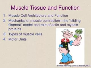
Muscle Cell Function
Muscle Cell Function. Vertebrate Anatomy – Ch. 9 AP Biology – Ch. 49. Functions of Muscle. Maintain body posture Stabilize joints Provide mobility Generate heat. Three Types of Muscle. Smooth muscle Found in the organs Stomach, intestines, etc. Involuntary Unconscious control.

Muscle Cell Function
E N D
Presentation Transcript
Muscle Cell Function Vertebrate Anatomy – Ch. 9 AP Biology – Ch. 49
Functions of Muscle • Maintain body posture • Stabilize joints • Provide mobility • Generate heat
Three Types of Muscle • Smooth muscle • Found in the organs • Stomach, intestines, etc. • Involuntary • Unconscious control
Three Types of Muscle • Cardiac Muscle • Found in the heart only • Involuntary • Has it’s own specialized electrical system allowing the heart to contract and relax on its own – no stimulus from the nervous system required • A single cardiac muscle cell, if left without input, will contract rhythmically at a steady rate; if two cardiac muscle cells are in contact, whichever one contracts first will stimulate the other to contract, and so on.
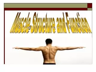
Skeletal Muscle • Associated with the bones • Allows for movement • Appears Striated (banding pattern) • VOLUNTARY
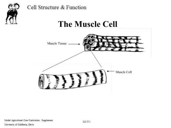
Long, cylindrical cell Produced by union of many embryonic cells Leads to huge cells Up to 12 inches long 10x larger in diameter than avg. cell Diagram Muscle cell structure
Muscle cell structure • Many nuclei in each fiber • Arranged just below the plasma membrane • Again, indicates fusion of many cells • Nuclei pushed to periphery to make more room for unified contracting fibers • Diagram
Muscle cell structure • Plasma membrane – sarcolemma • Cytoplasm – sarcoplasm • Lots of stored glycogen • Oxygen binding protein called myoglobin • Likehemoglobin
Muscle fiber - close up • Each muscle fiber contains a large number of rod-like myofibrils that run in parallel fashion and extend the entire length of the cell. • Myofibrils are densely packed.
Muscle fiber – close up • Myofibrils appear banded • Banding is due to two types of smaller fibers • Thick filaments • Thin filaments
Myofibrils – thick and thin filaments • Thick filament • Myosin • Thin filament • Actin
Thick filament structure • Myosin • Myosin heads • Myosin heads can move and “stick to” actin at certain locations on the actin fiber
Thin filament structure • Actin • Twisted strings of pearls • Possess binding sites • Locations where myosin heads can bind • These remain hidden when the muscle is NOT contracting • Troponin and Tropomyosin • Molecules associated with the actin filaments • Troponin • Calcium binding sites on actin filaments • Tropomyosin • “rope” molecule that covers and hides myosin binding sites
The Sarcomere • Z-line – anchor points for actin filaments • Sarcomere – from z-line to z-line • The sarcomere is the unit of contraction.
Interaction of Thick and Thin filaments – muscle contraction • Myosin head bound to ATP • In low energy state • Energy from ATP cannot be released until ATP is broken into ADP and Pi
Interaction of Thick and Thin filaments – muscle contraction • Myosin head hydrolyzes (breaks down) ATP to ADP and Pi. • Myosin head moves to its high energy position.
Interaction of Thick and Thin filaments – muscle contraction • Myosin head binds to actin forming a cross bridge • This occurs as long as the myosin binding sites on the actin filaments have been uncovered • Remember these were hiddin by the tropomyosin molecule
Interaction of Thick and Thin filaments – muscle contraction • Once the myosin head binds to actin, it releases ADP and Pi • Once ADP and Pi are released, myosin head returns to Low Energy State • REMEMBER: It is still hooked to the actin • THIS SLIDES THE THIN FILAMENT
Interaction of Thick and Thin filaments – muscle contraction • A new ATP binds to the myosin head • This causes a shape change in the myosin head that releases the myosin head from the actin molecule
How it looks… • Contraction of a sarcomere
But that’s not all…. • Remember • Tropomyosin • Troponin • Calcium • Where do these chemicals fit it in? • CONTROL of the muscle contraction
Control of muscle contraction • Tropomyosin covers myosin head binding sites on actin filaments • Troponin – calcium binding sites
Control of muscle contraction • Calcium binds to troponin • Troponin bound by Calcium moves tropomyosin • Reveals myosin head binding sites • Allows binding to occur
Where does Calcium come from and WHY? • Calcium is stored in the Sarcoplasmic reticulum (fancy ER) • Nerve impulse releases calcium from SR into sarcoplasm (cytoplasm • Nerve impulse travels across synapse to sarcolemma • Down T-tubules • To SR • When nerve impulse reaches SR, Calcium is released • Allows muscle contraction to occur, ONLY when nerve impulse has told it to do so.
Links to animations • How Muscles Work • Sarcomere shortening • Sliding Filament Theory (includes nerve stimulation)