Download
1 / 84
870 likes | 941 Vues
Explore animal form, size, and function, tissue structure, and thermoregulation in this comprehensive guide. Learn about nutrition stages, animal diversity, and symmetry patterns like bilateral and radial symmetry.

E N D
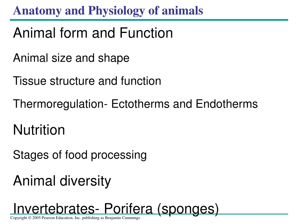
Anatomy and Physiology of animals Animal form and Function Animal size and shape Tissue structure and function Thermoregulation- Ectotherms and Endotherms Nutrition Stages of food processing Animal diversity Invertebrates- Porifera (sponges)
Text devoted to animals • Except for Chapter 8 Animal like protists (Amoeba and Paramecium) • The inclusion of protozoa is part of a tradition • Once considered a phylum (Protozoa) in the animal kingdom
Pattern of Organization • Symmetry • Asymmetry • Radial symmetry • Bilateral symmetry
Figure 7.7 Asymmetry red encrusting sponge asymmetrical
Figure 7.8 Radial symmetry tube coral pulp
Bilateral animals • Bilateral symmetry = important evolutionary advancement • Important for active, directed movement • Anterior, posterior ends • One side of body kept up (dorsal) vs. down (ventral)
Directed movement evolved with anterior sense organscephalization Cephalization • specialization of sense organs in head end of animals
Bilateral Symmetry • Divided along sagittal plane into two mirror images • sagittal= divides bilateral organisms into right and left halves
Anterior= head end • Posterior= tail end • Dorsal= back side • Ventral= belly side
Symmetry, fig. 7.9 • Median= sagittal
Other Patterns of Organization may reflect evolutionary trends • Unicellular (cytoplasmic)- organisms consist of single cells or cellular aggregates, • provide functions of locomotion, food acquisition, digestion, water and ion regulation, sensory perception and reproduction in a single cell. • Cellular aggregates consist of loose association, cells that exhibit little interdependence, cooperation, or coordination of function • Some cells may be specialized for reproduction, nutritive or structural function
Diploblastic Organization • Cells are organized into tissues in most animal phyla • Body parts are organized into layers derived from two embryonic tissue layers. • Ectoderm- Gr. ektos, outside + derm, skin gives rise to the epidermis the outer layer of the body wall • Endoderm- Gr. Endo, within, gives rise to the gastrodermis that lines the gut
Mesoglea- between the ecto and endo and may or may not contain cells • Derived from ecto and/or endo • Cells form middle layer (mesenchyme) • Layers are functionally inderdependent, yet cooperate showing tissue level organization i.e. feeding movements of Hydra or swimming movements of a jellyfish
The Triploblastic (treis, three +blaste, sprout) • Animals described in chapters 10-22 • Tissues derived from three embryological layers • Ectoderm- outer layer • Endoderm- lines the gut • Mesoderm- meso, middle, Third layer between Ecto and Endo • Give rise to supportive cells
Most have an organ system level of organization • Usually bilaterally symmetrical or evolved from bilateral ancestors • Organized into several groups based on the presence or absence of body cavity and for those that posses one, the kind of body cavity present. • Body cavity- fluid filled space in which the internal organs can be suspended and separated from the body wall
Body cavities are advantageous • Provide more room for organ development • Provide more surface area for diffusion of gases, nutrients, and waste into and out of organs • Provide area for storage • Often act as hydrostatic skeletons (supportive yet flexible) • Provide a vehicle for eliminating wastes and reproductive products from the body • Facilitate increase in body size
What does acoelomate mean? • No coelom
Acoelomate a, without+ kilos, hollow • Mesoderm relatively solid mass • No cavity formed between ecto and endo • These cells within mesoderm often called parenchymal cells • Parenchymal cells not speciallized for a particular fnc.
Earthworm What’s a coelom? • coelom= • true body cavity • Fluid-filled • lined by mesoderm-derived epithelium
Acoelomates lack a true body cavity • Solid body • no cavity b/w the digestive tract and outer body wall
Do these questions now… • Think about aceolomate bilateral animals: • To what domain do they belong • “ ” kingdom ” ” ” • What phyla include these organisms • What is bilateral symmetry, and why was it an important evolutionary advantage movie
Acoelomate Bilateral Animals • Consist of phyla: • Phylum Platyhelminthes • Phylum Nemertea • Others… flatworm
Reproductive and osmoregulatory systems Acoelomate Bilateral Animals • Simplest organisms to have bilateral symmetry • Triploblastic • Lack a coelom • Organ-system level of organization • Cephalization • Elongated, without appendages
Reproductive and osmoregulatory systems Acoelomate Bilateral Animals • Simplest organisms to have bilateral symmetry • Triploblastic • Lack a coelom • Organ-system level of organization • Cephalization • Elongated, without appendages
Triploblastic Pseudocoelomate pseudes, false • Body cavity not entirely lined by mesoderm • No muscle or connective tissue associated with gut • No mesodermal
The Triploblastic Coelomate Pattern • Coelom is a body cavity completely surrounded by mesoderm • Peritoneum- mesodermal sheet that lines the inner body wall and serosa (outer covering of visceral organs) • Having mesodermally derived tissue (muscle, connective tissue) enhances the function of all internal body systems.
Figure 7.3 Groups traced to separate ancestors All descendants of a single ancestor Includes some but not all of a members of a lineage Fig 7.3 Evolutionary groups
Fig 7.4 Vertebrate Phylogenetic tree depicts the degree of divergence from a common ancestor
Figure 7.5 Fig 7.5 Interpreting Cladograms Five taxa (1-5) and characteristics (A-H) Symplesiomorphies- common characters in a group
Figure 40.1 • The comparative study of animals • Reveals that form and function are closely correlated
Physical laws and the environment constrain animal size and shape • Physical laws and the need to exchange materials with the environment • Place certain limits on the range of animal forms
Physical Laws and Animal Form • Evolutionary convergence • Reflects different species’ independent adaptation to a similar environmental challenge (a) Tuna (b) Shark (c) Penguin (d) Dolphin (e) Seal Figure 40.2a–e
Exchange with the Environment • An animal’s size and shape • Have a direct effect on how the animal exchanges energy and materials with its surroundings • Exchange with the environment occurs as substances dissolved in the aqueous medium • Diffuse and are transported across the cells’ plasma membranes
Diffusion (a) Single cell • A single-celled protist living in water • Has a sufficient surface area of plasma membrane to service its entire volume of cytoplasm
Multicellular organisms with a sac body plan • Have body walls that are only two cells thick, facilitating diffusion of materials Mouth Gastrovascular cavity Diffusion Diffusion (b) Two cell layers
Organisms with more complex body plansHave highly folded internal surfaces specialized for exchanging materials External environment Food CO2 O2 Mouth Animal body Respiratory system Blood 50 µm 0.5 cm A microscopic view of the lung reveals that it is much more spongelike than balloonlike. This construction provides an expansive wet surface for gas exchange with the environment (SEM). Cells Heart Nutrients Circulatory system 10 µm Interstitial fluid Digestive system Excretory system The lining of the small intestine, a diges- tive organ, is elaborated with fingerlike projections that expand the surface area for nutrient absorption (cross-section, SEM). Inside a kidney is a mass of microscopic tubules that exhange chemicals with blood flowing through a web of tiny vessels called capillaries (SEM). Anus Unabsorbed matter (feces) Metabolic waste products (urine)
Animal form and function are correlated at all levels of organization • Animals are composed of cells • Groups of cells with a common structure and function • Make up tissues • Different tissues make up organs • Which together make up organ systems
Tissue Structure and Function • Different types of tissues • Have different structures that are suited to their functions • Tissues are classified into four main categories • Epithelial, connective, muscle, and nervous
Epithelial Tissue • Epithelial tissue • Covers the outside of the body and lines organs and cavities within the body • Contains cells that are closely joined
Epithelial tissue EPITHELIAL TISSUE Columnar epithelia, which have cells with relatively large cytoplasmic volumes, are often located where secretion or active absorption of substances is an important function. A simple columnar epithelium A stratified columnar epithelium A pseudostratified ciliated columnar epithelium Stratified squamous epithelia Cuboidal epithelia Simple squamous epithelia Basement membrane 40 µm
Connective Tissue • Connective tissue • Functions mainly to bind and support other tissues • Contains sparsely packed cells scattered throughout an extracellular matrix
CONNECTIVE TISSUE • Connective tissue 100 µm Chondrocytes Collagenous fiber Chondroitin sulfate Elastic fiber 100 µm Cartilage Loose connective tissue Adipose tissue Fibrous connective tissue Fat droplets Nuclei 150 µm 30 µm Blood Bone Central canal Red blood cells White blood cell Osteon Plasma 700 µm 55 µm
Muscle Tissue • Muscle tissue • Is composed of long cells called muscle fibers capable of contracting in response to nerve signals • Is divided in the vertebrate body into three types: skeletal, cardiac, and smooth


![[PDF READ ONLINE] Anatomy and Physiology of Farm Animals](https://cdn7.slideserve.com/12501546/slide1-dt.jpg)