Gastric T umours
290 likes | 311 Vues
This article provides an overview of different types of gastric tumours, including gastric polyps, carcinoids, and carcinomas. It covers their etiology, molecular biology, pathology, clinical features, and management options. The prognosis of gastric tumours is also discussed.

Gastric T umours
E N D
Presentation Transcript
Gastric Polyps • 1. Metaplastic- due to H. pylori infection, responds to eradication treatment. • 2. Inflammatory polyps • 3.Fundic polyps- due to PPI drugs or as a part of FAP syndrome. • 4. Adenomas- about 10% become malignant • 5. Carcinoid tumours of stomach
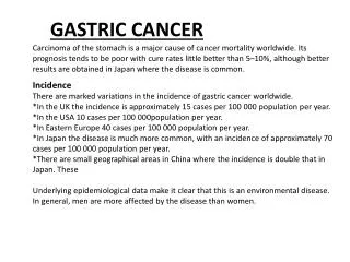
Carcinoma of Stomach • Cancer with poor prognosis unless detected and treated in early stages • The only curative modality of treatment is surgical • Incidence- 40/ 100,000 per year. In Japan 70 • More common in males, elderly
Location • More common in distal stomach in patients from lower socio-economic background and proximal stomach in patients from upper socio- economic group • Carcinomas from body and distal stomach are often associated with H. pylori infection unlike proximal gastric or Gastroesophageal (GE) junction tumours
Etiology Multifactorial • H. pylori • Pernicious anemia & gastric atrophy • Gastric polyps- adenomas • Previous gastric surgeries • Smoking • Diet- high salt intake, deficient antioxidants, N-nitroso compounds • Obesity • Genetic.
Molecular Biology of gastric ca • Less well understood than colorectal carcinoma. • Intestinal type-50% of patients have mutation or heterozygosity in APC gene. 30% patients have beta catenin mutation. Loss of heterozygosity at bcl-2 gene(inhibition of apoptosis)
Diffuse type- 50% patients have E- cadherin mutation. Rarely mutations in APC gene. • Both intestinal and diffuse type- 15% may have microsatellite instability(MSI – HNPCC/ Lynch syndrome) which is a DNA replication error. 30% patients have inactivation of p53 gene. Overexpression of growth factor receptors- c-Met, k- Sam,c-ErB2. Overexpression of transforming growth factor alpha, epidermal growth factor(EGF), vascular endothelial growth factor(VEGF)
Pathology • Lauren classification- 1. Intestinal gastric carcinoma: polypoidaltumours. Arise from intestinal metaplasia. Better prognosis 2. Diffuse gastric carcinoma- diffuse infiltration of gastric wall without forming any localised lesions 3. Mixed type- • Early Gastric Carcinoma/ Advanced Gastric Ca
Early gastric carcinoma- malignant cell infiltration confined to mucosa and submucosa with or without lymph node metastasis. Protruding , superficial, excavated ( Japanese). Curable. 5 year survival is upto 90%. In Japan 1/3 of stomach cancers are early gastric carcinomas.
Advanced gastric cancers- malignant cells infiltrate muscularis layer. Bormann’s classification- Type I, II, III, IV.
Spread -Distant spread is not common before local lymph node involvement. • Direct spread- muscularis layer, serosa, adjacent organs like pancreas, colon, liver. • Lymphatic spread- by permeation and embolisation. Supraclavicular lymph node involvement in advanced stage- Troisier’s sign. • Haematogenous- liver, lung, bone.
Transperitoneal spread- rectovesical/ rectovaginal pouch deposit- Blumer’s shelf. Ovaries in premenopausal women- Krukenberg’stumour. Umbilical deposits- Sister Mary Joseph nodules. Ascites.
Clinical Features • Early gastric carcinoma- nonspecific symptoms, dyspepsia. May be detected during gastroscopy for screening (Japan) or dyspepsia. • Exclude gastric cancer in all patients before PPI.
Advanced stage- • Early satiety, bloating, abdominal distention, vomiting, haematemesis, melaena or iron deficiency anemia • Obstruction to GE junction causes dysphagia, epigastric fullness, vomiting • Pyloric obstruction- features of gastric outlet obstruction. Alkalosis is mild or absent • Thrombophlebitis (Trousseau’s sign), deep vein thrombosis.
Management • Gastroscopy, biopsy • Hb , PCV • Serum electrolytes • Blood urea , serum creatinine • USG abdomen • CT scan abdomen • PET- CT scan • Chest X ray • Pre operative laparoscopy
If not operable- palliative procedures- palliative resection, palliative gastro-jejunostomy, Devine’s gastric exclusion, endoscopic stenting across the gastric tumour.
Chemotherapy- pre-op chemotherapy is effective. • Combination of Epirubicin, Cisplatinum, 5 Fluorouracil/ Capacitabine • Taxotere, Oxaliplatin • Trastuzumab( Herceptin)- in advanced, metastatic Her 2 receptor positive gastric cancers • Radiotherapy has no significant role.
Prognosis • Japan- 5 year survival is 50-70%. • West- 25-30%