
The Heart
The Heart. http://www.annerpino.com/2003/heart.jpg. Heart Anatomy. Heart Anatomy. Bicuspid / Mitral Valve. Pulmonary Artery / Trunk. Pulmonary Vein. Left Atrium. Left Ventricle. Aortic Valve. Aorta. Superior Vena Cava. Inferior Vena Cava. Pulmonary Valve. Tricuspid Valve.
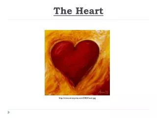

The Heart
E N D
Presentation Transcript
The Heart http://www.annerpino.com/2003/heart.jpg
Heart Anatomy Bicuspid / Mitral Valve Pulmonary Artery / Trunk Pulmonary Vein Left Atrium Left Ventricle Aortic Valve Aorta Superior Vena Cava Inferior Vena Cava Pulmonary Valve Tricuspid Valve Right Ventricle Right Atrium
Blood Pressure Diastole: • When atria are relaxed (i.e. filled with blood) Systole: • Atria push blood into ventricles • Ventricles contract and push blood into arteries • When AV valves shut – LUBB • Ventricles relax – DUBB Written as: DIASTOLIC pressure (mm Hg) SYSTOLIC pressure (mm Hg)
How do we take Blood Pressure? • Need to use a stethoscope • Normal systolic should be 120 mm Hg • Normal diastole should be 80 mm Hg To measure: • Tighten the cuff…as soon as you release you should hear a lubb sound – measure pressure (systolic – should be 120 mm Hg) • Then deflate until sound disappears – check pressure (diastole – should be 80mm Hg) Sphygmomanometer
Heart Valves • 4 valves ensure that blood • flows in the proper direction • 1) Tricuspid (“right atrioventricular/AV-valve”) • - b/w RA & RV • 2) Bicuspid/Mitral (“left AV-valve”) • – b/w LA & LV • Pulmonary semilunar valve • - b/w RV & pulmonary trunk • Aortic semilunar valve • - b/w LV & aorta • http://www.nhlbi.nih.gov/health/dci/Diseases/hhw/hhw_electrical.html ** CHORDAE TENDINAE (“heart strings”) - Fibrous connective tissue inside ventricles that stabilize the AV valves
Coronary Circulation • supply oxygenated, nutrient-rich blood to the muscles of the heart (cardiac muscle) • disease of this “mini-circulatory system” will lead to death of heart tissue • unfortunately, cardiac muscle is not able to regenerate • if too much tissue dies, the heart cannot pump effectively = heart attack


