Download
1 / 26
260 likes | 613 Vues
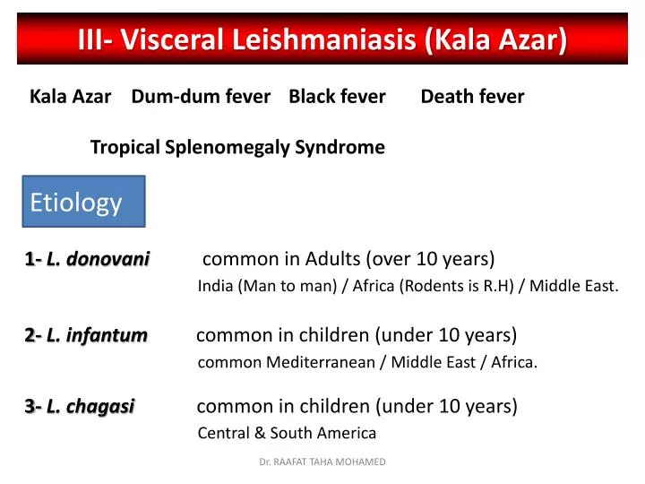
III- Visceral Leishmaniasis (Kala Azar). Kala Azar. Dum-dum fever. Black fever. Death fever. Tropical Splenomegaly Syndrome. Etiology. 1- L. donovani common in Adults (over 10 years) India (Man to man) / Africa (Rodents is R.H) / Middle East. .

E N D
III- Visceral Leishmaniasis (Kala Azar) Kala Azar Dum-dum fever Black fever Death fever Tropical Splenomegaly Syndrome Etiology 1- L. donovani common in Adults (over 10 years) India (Man to man) / Africa (Rodents is R.H) / Middle East. 2- L. infantum common in children (under 10 years) common Mediterranean / Middle East / Africa. 3- L. chagasi common in children (under 10 years) Central & South America Dr. RAAFAT TAHA MOHAMED
III- Visceral Leishmaniasis (Kala Azar) Epidemiology * Cases of V.L. recorded annually are about 40.000. * Annual death range from 5.000 -10.000. * The Disease is endemic in Mediterranean area & became indicator for AIDS patients in Spain. * The Disease is mainly Zoonotic among dogs & Rodents. * In India & Bangladesh it is Anthroponotic. * Endemicity is determined by close contact between Reservoir Host & Vectors . Dr. RAAFAT TAHA MOHAMED
Visceral Leishmaniasis Dr. RAAFAT TAHA MOHAMED
Pathogenesis of Visceral Leishmaniasis 1-Leishmanioma: Small papule --- Nodule (rarely Ulcerate) ----- Amastigote within skin macrophages. • N.B: - Effective cellular immune response controls dissemination of infection. • Malnutrition & Immunosuppression --- contribute in disease development. • V.L. is an immunosuppressive disease. • Children & young Adults are more susceptible to the infection. • Immunocompromized develop highly fatal severely progressive disease. Dr. RAAFAT TAHA MOHAMED
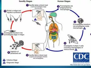
Pathogenesis of Visceral Leishmaniasis 2- Incubation period: several months to years, now it can be few days 3- Infection spread from primary skin lesionhaematogenously to organs R.E.S. (BONE MARROW – LYMPHOID TISSUE – SPLEEN – LIVER – G.I.T …etc. 4-Amastigote multiply intracellularly granulomatousreactionHyperplasia & Hypertrophy of affected organ. Progressive disease may lead to the followings: 1-Bone marrow suppressionPANCYTOPENIA ( Anemia – Leucopenia /Neutropenia “tendency to bacterial infection” – Thrombocytopenia “bleeding tendency”). 2- Massive Splemomegalyearly destruction & short life span of R.B.Cs Anemia. Dr. RAAFAT TAHA MOHAMED
Pathogenesis of Visceral Leishmaniasis Progressive disease may lead to the followings: 3- Secondary bacterial infection ( due to Neutropenia / compromised immunity / malnutrition) Otitis media – Pneumonia - Septicemia 4- Liver affection reversed A/G ratio Hypoalbuminaemia Oedema. 5- Immune complexes mild glomerulonephritis (deposition of C.I.C.in glomerular capillaries)Nephrotic syndrome may lead to death. Dr. RAAFAT TAHA MOHAMED
Pathogenesis ofVisceral Leishmaniasis Leishmaniomais rarely seen at the site of bite Via blood stream to Taken by Promastigotes Amastigotes Reticulo-endothelial cells present in human viscera RBCs Liver: hepatomegaly Spleen: splenomegaly WBCs marrow Lymph nodes: enlarged Bone marrow: Anaemia, leukopenia, thrombocytopenia Platelets Reversal of albumin / globulin ratio Intestinal mucosa: diarrhoea & dysentery Dr. RAAFAT TAHA MOHAMED
Clinical picture of Visceral Leishmaniasis Asymptomatic “ most common” Symptomaticinfections may lead to the following: 1- Leishmanioma (Primary skin lesion). 2- Fever: irregular – prolonged – intermittent (2-3 peaks daily). dromedary fever 3- Splenomegaly: progressively enlarged /soft / tender 4- Hepatomegaly: progressively enlarged /soft / tender 5- Lymphadenopathy. 6- Jaundice. 7- Skin is darkly Pigmented at face , hands & feets 8- Diarrhea or Dysentery. 9- Weight loss. Dr. RAAFAT TAHA MOHAMED
Clinical Picture of Visceral Leishmaniasis 1- Fever: intermittent with double daily rise. Also called Dum-dum fever (Dum-dum is a town in Calcutta in India) 2- hepatosplenomegaly NWVLhepatosplenomegaly OWVLhepatosplenomegaly Dr. RAAFAT TAHA MOHAMED
Clinical Picture of Visceral Leishmaniasis 3- Skin changes Disturbance of pigmentation occurs Dark pigmentation or depigmentation (butterfly pigmentation) (also called Kala azarكلمة باللغة الهنديةmeans black fever) 4- Post kalaazar dermal leishmanoid (PKDL) Skin nodules. Persistent allergic reaction to parasite antigens Seen in two conditions 1- Spontaneous arrest of the disease 2- Incomplete antimony treatment 5- Weight loss, emaciation, death from inter-current infection (cancrumoris, pulmonary, gastrointestinal infections). Leishmania causes suppression of cell-mediated immunity Dr. RAAFAT TAHA MOHAMED
Post KALA-AZAR Dermal Leishmaniasis (P.K.D.L) * About 15-20% of V.L. patients develop P.K.D.L in 1-5 years post infection * Skin lesion develops after apparent cure or shortly after or during treatment • Lesions appear as follows: • (A)- Multiple depigmented macules /papules/nodules. • (B)- Common in face resemble “Lepromatous leprosy” – • Limbs – upper trunk. • (C)- Chronic persistent lesion rarely ulcerate. Dr. RAAFAT TAHA MOHAMED
Skin changes in visceral leishmaniasis Butterfly pigmentation Post Kala-Azar dermal leishmanoid Usually occur in patients from the old world Depigmented areas Dr. RAAFAT TAHA MOHAMED
Areas where Visceral Leishmaniasis exists L.infantum L.donovani Brazil L.chagasi New world visceral leishmaniasis Old world visceral leishmaniasis Dr. RAAFAT TAHA MOHAMED
Diagnosis of C. Leishmaniasis V. Leishmaniasis Amastigotes Clinically: Clinically: Necrotic tissue Ulcer with sharp cut indurated margin Fever, hepatosplenomegaly Microscopy: Microscopy: To detect amastigotesin blood, liver, spleen, lymph node, bone marrow To detect amastigotesat the edge of the ulcer by aspiration or biopsy Culture: Culture: To detect promastigotes To detect promastigotes Animal inoculation Montenegro test Montenegro test +ve after successful treatment ˃ 95% Dr. RAAFAT TAHA MOHAMED Serological tests Serological tests
Aspiration and biopsy from the ulcer Aspiration Scrape or take biopsy Leishmaniaamastigotes (Giemsa stained) Dr. RAAFAT TAHA MOHAMED
Aspiration and biopsy from Bone marrow Bone marrow Amastigote Bone marrow aspiration Supportive Laboratory Findings: 1- Reversal A/G ratio. 2- Hypergammaglobinaemia. 3- Anemia. 4- Neutropenia. 5- Thrombocytopenia. Dr. RAAFAT TAHA MOHAMED
Diagnosis (cont.) • Promastigotes in rosettesin a culture of an orient sore on N.N.N. medium (Giemsa stain). Montenegro “Leishmanin” Test: 1-Detects Delayed type Hypersensitivity. 2- -veduring acute stage of V.L. 3- +veafter cure. 4- Negative for D.C.L. (suppressed immunity). 5- In Endemic areas, high % of population are positive. Dr. RAAFAT TAHA MOHAMED
Diagnosis (cont.) Immunological Diagnosis: • Specific serologic tests: Direct Agglutination Test (DAT), ELISA, IFAT • Non specific detection of hypergammaglobulinaemia by formaldehyde (formol-gel) test or by electrophoresis. Dr. RAAFAT TAHA MOHAMED
Buffy Coat Method A technique used for collection of parasite from blood sample Plasma WBCs, platelets, parasite Buffy coat RBCs Centrifuge Patient’s blood Dr. RAAFAT TAHA MOHAMED
Treatment Cutaneous leishmaniasis Visceral leishmaniasis Antimony sodium gluconate (Pentostam) Given intramuscularly Treatment of ulcer Surgical excision Curettage Physical Heat, freezing 2% chlopromazine & clortrimazole Chemical I.D. injection of interferon gamma around the lesion to promote healing of the ulcer Dr. RAAFAT TAHA MOHAMED
Control of Leishmaniasis • Treatment of Patients • Protection: by using Wire screens, repellents & mosquito nets • Control of sandfly vector • Vaccination in endemic areas gives long lasting immunity Dr. RAAFAT TAHA MOHAMED
Case A female patient went to dermatology clinic suffering from skin lesion. On examination, the doctor noticed a skin ulcer with sharp edge and indurated margin The patient gave a history of an arthropod bite. a- What is your provisional diagnosis? A case of Cutaneous leishmaniasis b- How can you confirm your diagnosis? By microscopy, culture, skin test or serological tests. Amastigotes c- How can you manage such condition? Pentostam I.M., Physical, chemical methods, ID injection of interferon gamma of ulcer Dr. RAAFAT TAHA MOHAMED
Case A young Egyptian arriving from Jordan where he was working as a laborer and living in campus. He has a chronic ulcer on his arm with sharp-cut edge that resist treatment by known antibiotics. a- What is your provisional diagnosis? Cutaneous leishmaniasis. b- How can you confirm your diagnosis? By microscopy, culture, skin test or serological tests. c- How can you manage such case? Pentostam I.M. and surgical or chemical treatment for ulcer. c- How can you control such infection? Treat patients, protect the healthy, control of sandfly. Dr. RAAFAT TAHA MOHAMED
Compare between • Diagnosis of Cutaneous and Visceral leishmaniasis. Detection ofamastigotesfrom edge of the ulcer Detection ofamastigotesin blood, liver, spleen, lymph node, bone marrow • Ulcer produced by Montenegro test positive in 95% of cases Montenegro test positive after successful treatment L.tropica L.major Dry, single Wet, multiple chronic acute In urban areas In rural areas Dr. RAAFAT TAHA MOHAMED
MCQ Stained smears from organs in kala-azar show: a- Epimastigote form c- Amastigote form b- Promastigote form d- Trypomastigote form To diagnose cutaneousleishmaniasis, biopsy is better taken from: a- base of the ulcer c- lymph node draining the ulcer b- edge of the ulcer d- skin around the ulcer The following arthropod transmits visceral leishmaniasis: a- Culicoides c- Simulium b- Chrysops d- Phlebotomus Dr. RAAFAT TAHA MOHAMED
State true or false • To diagnose cutaneousleishmaniasis a biopsy is better taken from the base of the ulcer. False A biopsy is better taken from the edge of the ulcer where Leishmaniaamastigotes exist, the base of ulcer contains only necrotic tissue • Amastigote forms of Leishmania occur in culture. Promastigote forms of Leishmania occur in culture False Give reason Leishmania parasites are present in small number in patient's blood. Leishmania parasites are taken by the reticuloendothelial cells of viscera Dr. RAAFAT TAHA MOHAMED