Chapter 14 The Central Nervous System
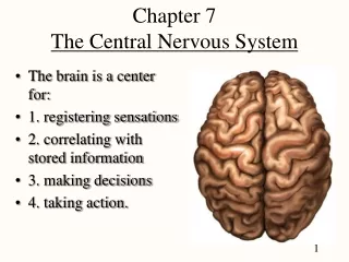
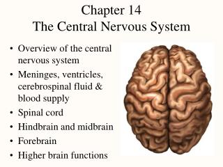
Chapter 14 The Central Nervous System. Overview of the central nervous system Meninges, ventricles, cerebrospinal fluid & blood supply Spinal cord Hindbrain and midbrain Forebrain Higher brain functions. Brain Description. Brain weighs 3 to 3.5 pounds

Chapter 14 The Central Nervous System
E N D
Presentation Transcript
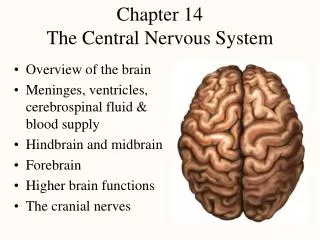
Chapter 14The Central Nervous System • Overview of the central nervous system • Meninges, ventricles, cerebrospinal fluid & blood supply • Spinal cord • Hindbrain and midbrain • Forebrain • Higher brain functions
Brain Description • Brain weighs 3 to 3.5 pounds • Major portions of the brain--brainstem, cerebrum, and cerebellum • cerebrum is 83% of brain volume; cerebellum contains 50% of the neurons
Brain Longitudinal fissure separates 2 cerebral hemispheres. Central sulcus separates frontal and parietal lobe.
Embryonic Development • Nervous system develops from ectoderm • by 3rd week, neural plate becomes a groove with neural folds along each side • by 4th week, neural folds join to form neural tube • lumen of the neural tube develops into central canal of spinal cord & ventricles of the brain • cells along the margin of the neural groove is called the neural crest • develop into sensory and sympathetic neurons & schwann cells • by 4th week, neural tube exhibits 3 anterior dilations
Brain Development • 4th week • forebrain • midbrain • hindbrain • 5th week • telencephalon • diencephalon • mesencephalon • metencephalon • myelencephalon
Meninges • Dura mater -- outermost, tough membrane • outer periosteal layer against bone • where separated from inner meningeal layer forms dural venous sinuses draining blood from brain • supportive structures formed by dura mater • falx cerebri, falx cerebelli and tentorium cerebelli • epidural space filled with fat in lower back region • epidural anaesthesia during childbirth • Arachnoid mater is spider web filamentous layer • Pia mater is a thin vascular layer adherent to contours of brain
Ventricles and Cerebrospinal Fluid • Internal chambers within the CNS • lateral ventricles found inside cerebral hemispheres • third ventricle is single vertical space under corpus callosum • cerebral aqueduct runs through midbrain • fourth ventricle is small chamber between pons & cerebellum • central canal runs down through spinal cord • Lined with ependymal cells and containing choroid plexus of capillaries that produce CSF
Cerebrospinal Fluid • Clear liquid fills ventricles and canals & bathes its external surface (in subarachnoid space) • Brain produces & absorbs about 500 ml/day • filtration of blood through choroid plexus • has more Na+ & Cl- but less K+ & Ca+2 than plasma • Functions • buoyancy -- floats brain so it neutrally buoyant • protection -- cushions from hitting inside of skull • chemical stability -- rinses away wastes • Escapes from 4th ventricle to surround the brain • Absorbed by arachnoid villi into venous sinus
Blood-Brain and Blood-CSF Barriers • Blood-brain barrier is tightly joined endothelium • permeable to lipid-soluble materials (alcohol, O2, CO2, nicotine and anesthetics) • administer drugs through nasal sprays • circumventricular organs in 3rd & 4th ventricles at breaks in the barrier where blood has direct access • monitoring of glucose, pH, osmolarity & other variations • allows route for HIV virus to invade the brain • Blood-CSF barrier at choroid plexus is ependymal cells joined by tight junctions
Functions of the Spinal Cord • Conduction • bundles of fibers passing information up & down spinal cord • Locomotion • repetitive, coordinated actions of several muscle groups • central pattern generators are pools of neurons providing control of flexors and extensors (walking) • Reflexes • involuntary, stereotyped responses to stimuli • remove hand from hot stove
Anatomy of the Spinal Cord • Ropelike bundle of nerve tissue within the vertebral canal (thick as a finger) • vertebral column grows faster so in an adult the spinal cord only extends to L1 • 31 pairs of spinal nerves coming from cervical, thoracic, lumbar or sacral regions of the cord • named for level of vertebral column where nerves exit • Cervical & lumbar enlargements in cord • Medullary cone is tapered tip of spinal cord • Cauda equinae is L2 to S5 nerve roots resemble horse’s tail
Cross-Sectional Anatomy of the Spinal Cord • Central area of gray matter shaped like a butterfly and surrounded by white matter in 3 columns
Gray Matter • Pair of dorsal or posterior horns • dorsal root of spinal nerve is totally sensory fibers • Pair of ventral or anterior horns • ventral root of spinal nerve is totally motor fibers • Connected by gray commissure punctured by a central canal continuous above with 4th ventricle
White Matter • Bundles of myelinated axons that run up & down • Dorsal or posterior columns or funiculi • Lateral columns or funiculi • Anterior columns or funiculi • Each column is filled with tracts or fasciculi
Spinal Tracts • Ascending & descending tract head up or down while decussation means that the fibers cross sides • Contralateral means from the opposite side while ipsilateral means 2 regions on same side
CNS Ascending Pathway • Deep touch, vibration, limb movement & position (proprioception) • Fasciculus gracilis & cuneatus carry signals from arm & leg respectively • Decussation of 2nd order neuron in medulla
CNS Ascending Pathway 2 • Spinothalamic tract • Pain, pressure, temperature, light touch, tickle & itch • Decussation is in spinal cord
CNS Descending Pathway • Corticospinal tract • Motor signals from cerebral cortex for limb movements • Decussation in medulla forms lateral tract • anterior tract uncrossed • Tectospinal, reticulospinal & vestibulospinal tracts maintain posture & balance and provide reflex movements of the head
Medulla Oblongata • 3 cm extension of spinal cord • Ascending & descending nerve tracts • Nuclei of sensory & motor cranial nerves (IX, X, XI, and XII) • Cardiac center adjusts rate & force of heart beat • Vasomotor center adjusts blood vessel diameter • Respiratory centers control rate & depth of breathing • Reflex centers for coughing, sneezing, gagging, swallowing, vomiting, salivation, sweating, movements of tongue & head • Pyramids and olive visible on surface
Medulla and Pons Olive
Pons • Bulge in the brainstem, rostral to the medulla • Ascending sensory tracts • Descending motor tracts • Pathways in & out of cerebellum • Nuclei concerned with sleep, hearing, balance, taste, eye movements, facial expression, facial sensation, respiration, swallowing, bladder control & posture • cranial nerves V, VI, VII, and VIII
Cerebellum • Right & left hemispheres connected by vermis • Parallel surface folds called folia are gray matter • all of output comes from deep gray nuclei • large cells in single layer in cortex are purkinje cells synapse on deep nuclei
Cerebellum • Connected to brainstem by cerebellar peduncles • White matter (arbor vitae) visible in sagittal section • Sits atop the 4th ventricle
Midbrain, Cross Section • Mesencephalon • Central aqueduct • CN III and IV • eye movement • Cerebral peduncles hold corticospinal tract • Tegmentum connects to cerebellum & helps control fine movements through red nucleus • Substantia nigra sends inhibitory signals to basal ganglia & thalamus (degeneration leads to tremors and Parkinson disease)
Superior & Inferior Colliculus • Tectum (4 nuclei) called corpora quadrigemina • superior colliculus (tracking moving objects ) • inferior colliculus (reflex turning of head to sound)
Reticular Formation • Clusters of gray matter scattered throughout pons, midbrain & medulla • Regulate balance & posture • relaying information from eyes & ears to cerebellum • gaze centers allow you to track moving object • Includes cardiac & vasomotor centers • Origin of descending analgesic pathways • Regulates sleep & conscious attention • injury leads to irreversible coma
Thalamus • Oval mass of gray matter protruding into lateral ventricle (part of diencephalon) • Receives nearly all sensory information on its way to cerebral cortex • integrate & directs information to appropriate area • Interconnected to limbic system so involved in emotional & memory functions
Hypothalamus • Walls & floor of 3rd ventricle • Functions • hormone secretion & pituitary • autonomic NS control • thermoregulation (thermostat) • food & water intake (hunger & satiety) • sleep & circadian rhythms • memory (mammillary bodies) • emotional behavior
Cerebrum -- Gross Anatomy • Cerebral cortex is 3mm layer of gray matter with extensive folds to increase surface area ---- divided into lobes
Functions of Cerebrum Lobes • Frontal contains voluntary motor functions and areas for planning, mood, smell and social judgement • Parietal contains areas for sensory reception & integration of sensory information • Occipital is visual center of brain • Temporal contains areas for hearing, smell, learning, memory, emotional behavior • Insula is still little known
Tracts of Cerebral White Matter • Most of volume of cerebrum is white matter • Types of tracts • projection tracts • extend vertically from brain to spinal cord forming internal capsule • commissural tracts • cross from one hemisphere to the other • corpus callosum is wide band of white fiber tracts • anterior & posterior commissures are pencil-lead sized • association tracts • connect lobes & gyri of each hemisphere to each other
Cerebral Cortex • Surface layer of gray matter -- 3 mm thick • Neocortex (six-layered tissue) • newest part of the cortex (paleocortex & archicortex) • layers vary in thickness in different regions of brain • 2 types of cells • stellate cells • have dendrites projectingin all directions • pyramidal cells • have an axon that passes out of the area
Basal Nuclei • Masses of gray matter deep to cerebral cortex • Receive input from substantia nigra & motor cortex & send signals back to these regions • Involved in motor control & inhibition of tremors
Limbic System • Loop of cortical structures surrounding deep brain • amygdala, hippocampus, fornix & cingulate gyrus • Amydala important in emotions and hippocampus in memory -- rest are not sure
EEG and Brain Waves • Electroencephalogram records voltage changes from postsynaptic potentials in cerebral cortex • Differences in amplitude & frequency distinguish 4 types of brain waves
Brain Waves & Sleep • States of consciousness can be correlated with EEG • 4 types of brain waves • alpha occur when awake & resting with eyes closed • beta occur with eyes open performing mental tasks • theta occur during sleep or emotional stress • delta occur during deep sleep • Sleep is temporary state of unconsciousness • coma is state of unconsciousness with no possible arousal • reticular formation seems to regulate state of alertness • suprachiasmatic nucleus acts as biological clock to set our circadian rhythm of sleep and waking
Stages of Sleep • Non-REM sleep occurs in stages • 4 stages occurring in first 30 to 45 minutes of sleep • stage 1 is drifting sensation (would claim was not sleeping) • stage 2 still easily aroused • stage 3 vital signs change -- BP, pulse & breathing rates drop • reached in 20 minutes • stage 4 is deep sleep -- difficult to arouse • seems to have a restorative effect • REM sleep occurs about 5 times a night • rapid eye movements under the eyelids, vital signs increase, EEG resembles awake person, dreams and penile erections occur • may help sort & strengthen information from memory
Sleep Stages and Brain Waves • Brain waves change as we pass through 4 stages of sleep: alpha, to sleep spindles, to theta and finally to delta waves during deep sleep
Sleep Stages • Notice how REM sleep periods become longer and more frequent in the second half of the night
Cognition • Cognition is mental processes such as awareness, perception, thinking, knowledge & memory • 75% of brain is association areas where integration of sensory & motor information occurs • Examples of effects of brain lesions • parietal lobe -- contralateral neglect syndrome • temporal lobe -- agnosia (inability to recognize objects) or prosopagnosia (inability to recognize faces) • frontal lobe -- problems with personality (inability to plan & execute appropriate behavior)
Memory • Information management requires learning, memory & forgetting (eliminating the trivia) • pathological inability to forget have trouble with reading comprehension • anterograde amnesia -- can not store new data • retrograde amnesia -- can not remember old data • Hippocampus is important in organizing sensory & cognitive information into a memory • lesion to it causes inability to form new memories • Cerebellum helps learn motor skills • Amygdala important in emotional memory
Emotion • Prefrontal cortex controls how emotions are expressed (seat of judgement) • Emotions form in hypothalamus & amygdala • artificial stimulation produces fear, anger, pleasure, love, parental affection, etc. • electrode in median forebrain bundle in rat or human and a foot pedal • press all day to the exclusion of food (report a quiet, relaxed feeling) • Much of our behavior is learned by rewards and punishments or responses of others to them
Somesthetic Sensation • Somesthetic signals travel up gracile and cuneate fascicui and spinothalamic tracts of spinal cord • Somatosensory area is postcentral gyrus