The Digestive System Chapter 12
A&P 1 Tutor: Eleshia Howell. The Digestive System Chapter 12. All living organisms must obtain nutrients from their environment to sustain life. These substances are used as raw materials for synthesizing essential compounds or decomposed to provide the energy that cells require to function.

The Digestive System Chapter 12
E N D
Presentation Transcript
A&P 1 Tutor: Eleshia Howell The Digestive SystemChapter 12
All living organisms must obtain nutrients from their environment to sustain life. • These substances are used as raw materials for synthesizing essential compounds or decomposed to provide the energy that cells require to function. • In effect, the digestive system provides both the fuel that keeps all the body’s cells running and the building blocks needed for cell growth and repair.
The digestive system is the collective term used to describe the alimentary canal, it’s accessory organs and the variety of processes occurring at different levels in the canal. • The alimentary canal begins at the mouth, passes through the thorax, abdomen and pelvis, ending at the anus. • The digestive process gradually breaks down ingested food to a suitable form for absorption to occur ~ digestive enzymes.
The nutrients absorbed from the digestive system provide the ‘raw materials’ for manufacture of new cells, hormones, enzymes, and the energy required for cellular metabolism. • The activities of the digestive system can be grouped into 5 main categories: • Ingestion – eating & drinking • Propulsion – mixing & movement of contents • Digestion – mechanical and chemical breakdown • Absorption – fluid, nutrients, substances • Elimination – waste products
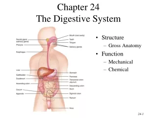
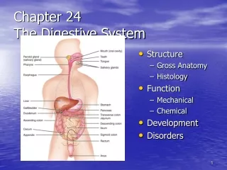
Organs of Digestive System Alimentary Canal – or gastro-intestinal tract (G.I.T) begins at the mouth and ends at the anus. Essentially a long tube through which food passes. • Mouth • Pharynx • Oesophagus • Stomach • Small intestine • Large intestine • Rectum and anal canal
Accessory Organs – various secretions are discharged into the alimentary tract, some by glands within the membranous lining of organs, and some by glands located outside the G.I.T. These ‘external’ structures are termed accessory organs of digestion and their secretions are provided via ducts to enter the G.I.T. • Salivary glands • Pancreas • Liver & biliary tract
The organs and glands are linked physiologically as well as anatomically ~ digestion and absorption occurs in stages, each stage being dependent upon the previous one(s).
Basic Structure of G.I.T • The layers of the walls of the alimentary canal follow a consistent pattern from the oesophagus onwards (not mouth & pharynx). • Modifications to this basic structure occur where specific function is required. • 4 layers to walls of alimentary tract: • Aventitia or serosa (outer) • Muscle layer • Submucosa • Mucosa (lining)
Adventitia or Serosa– • In the throrax, consists of loose fibrous tissue; in the abdomen the organs are covered by a serous membrane called peritoneum (the largest serous membrane in the body). • The peritoneum is a closed sac within the abdominal cavity, containing a small amount of serous fluid. It is richly supplied with blood & lymph vessels & contains numerous lymph nodes.
The peritoneum provides a physical barrier from the spread of local infection, but can also isolate local infection, eg appendicitis. • It has two layers – • Parietal – lining the abdominal wall • Visceral – covering the organs within abdominal and pelvic cavities. • The arrangement of the peritoneum is such that the organs are invaginated ~ secured in their own ‘pouch’ to keep them in place.
A storage of fat, known as the greater omentum, hangs in front of the abdominal organs, providing insulation and a long-term energy store. • The main blood vessels and nerves pass close to the abdominal wall and send branches to the organs between the folds of peritoneum. • The two layers of peritoneum are in contact, lubricated with serous fluid ~ potential cavity. In men, peritoneal cavity completely closed; in women there is allowance for the fallopian tubes & ovaries.
Muscle Layer – • Mainly consists of two layers of smooth (involuntary) muscle. • The muscle fibres in the outer layer are arranged longitudinally and the fibres of the inner layer form a circular wall of the tube. • Between the two layers are blood vessels, lymph vessels and network of sympathetic & parasympathetic nerves (mesenteric plexus) which serve the smooth muscle & blood vessels.
Contraction & relaxation of these muscles occurs in waves to push the contents of the digestive tract onwards. This is known as peristalsis, and is under control of SN and PSN. • Muscle contraction also mixes food with the digestive juices. • Movement through the G.I.T is controlled at various points by sphincters, which act as valves, preventing backflow and allowing time for digestion & absorption to occur.
Submucosa – • Consists of loose areolar connective tissue containing collagen and some elastic fibres, binding the muscle layer to the mucosa. • Arterioles, venules, capillaries and lymph vessels are contained within, as well as a submucosal plexus, also under SN and PSN supply.
Mucosa – • This consists of 3 layers of tissue ~ • Mucous membrane: columnar epithelium; provides protection, secretion & absorption. • Lamina propria: loose connective tissue, supporting blood vessels and lymph tissue to provide nourishment & protection • Muscularis mucosa: thin outer layer of smooth muscle, provides involutions eg glands, villi.
Mucous Membrane: • Parts of the G.I.T subject to wear and tear or mechanical injury consist of stratified squamous epithelial tissue with mucous secreting glands located just below the surface. • Areas where secretion of digestive juices and absorption occur consist of columnar epithelial tissue interspersed with goblet cells.
The goblet cells secrete mucous to lubricate the walls of the digestive tract and protect it from the damaging effect of digestive enzymes. • Other specialised glands or cells also lie below columnar cells to secrete substances such as saliva, gastric juice, intestinal juice, pancreatic juice and bile into the lumen of the tract to chemically break down food. • Lymphoid tissue is also situated below the tissue to protect against ingested microbes.
Nerve Supply • Alimentary canal and accessory organs are supplied by nerves from both divisions of the ANS ~ SNS & PSNS ~ providing antagonistic action, according to the body’s need at any particular time. • Digestion increased parasympathetic stimulation, eg peristalsis, secretions. • Sympathetic supply arises from numerous Thoracic & Lumbar spinal nerves (thoracic, abdominal, pelvic ganglia).
Fantastic Voyage... ...journey to the end of the alimentary canal!
The Mouth (oral cavity) • Lined throughout with mucous membrane, consisting of stratified squamous epithelium containing small, mucous secreting glands. • the palate forms the roof of the mouth; divided into anterior Hard palate (formed by maxilla & palatine bones), and posterior Soft palate (muscular soft tissue extending into the pharynx).
Hanging from the middle of the soft palate is the Uvula, from which 4 folds of tissue extend downwards to each side to form two membranous arches • Posterior – palatopharyngeal arch • Anterior – palatoglossal arch • Between the arches, on either side, are the tonsils.
TONGUE: • A voluntary muscular structure on the floor of the mouth, attached at its base to the Hyoid bone, and by a fold of its own mucous membrane covering called the frenulum. • The superior surface has numerous papillae containing sensory receptors for the sense of taste. • Blood supply – lingual branch of external carotid artery; lingual vein to internal jugular.
Nerve supply is from the hypoglossal (12th C.N), Facial (7th) and Glossopharyngeal (9th) and branches from mandibular nerves (branch from 5th) • Functions: • Chewing (mastication) • Swallowing (deglutition) • Speech • Taste
TEETH: • Are embedded in sockets of the alveolar ridge of the mandible and maxilla. • We are born with two sets ~ temporary (deciduous) and permanent ~ in immature form below the surface. • 20 x temporary teeth (10 each jaw), usually begin erupting from 6mths old; all should be present by 2yrs. • Permanent teeth (32) begin to replace temp’s from age 6; complete by 21yo.
Teeth have different functions, depending on their shape… • Incisors & canine ~ cutting, biting off pieces of food • Premolar & molar – grinding, chewing. • The centre of the tooth contains blood & lymph vessels & nerves, which pass through a small hole at the apex of each root of the tooth, into the tissue below. • Blood supply – maxillary arteries; internal jugular vein • Nerve – branches of trigeminal nerve (5Th)
SALIVARY GLANDS: • Release their secretions into ducts leading into the mouth • 3 main pairs – • Parotid - • Submandibular • Sublingual • Numerous other smaller glands scattered around the mouth • Saliva is the combined secretions from the glands & small mucous secreting glands of the oral mucosa.
About 1.5L of saliva is produced daily, consisting of: • Water • Mineral salts • A digestive enzyme = salivary amylase • Mucus • Lysozomes • Immunoglobulins • Blood-clotting factors • Secretion is under ANS control
Parasympathetic stimulation causes profuse secretion of watery saliva, relatively low enzyme & other organic substance content. • Sympathetic stimulation results in small amounts of organically rich saliva. • Reflex secretion occurs when there is food present and can be conditioned so that smell, sight and even thought of food stimulates production.
Functions of saliva include: • Chemical digestion of polysaccharides by amylase enzyme • Lubrication of food, forming a bolus ready for swallowing • Cleaning & lubricating the mouth, helping to prevent damage to mucous membranes by rough / abrasive food • Non-specific defence – lysozomes, antibodies & clotting factors • Taste – buds are stimulated only by chemical substances in solution
Pharynx • Divided into 3 sections: • Nasopharynx - respiration • Oropharynx • Laryngopharynx • Food passes from oral cavity into pharynx, then into oesophagus (continuous) • 3 layers of tissue: inner mucous membrane, middle connective tissue layer containing vessels & nerves, outer involuntary muscle layer (swallowing no longer voluntary from this point). Common to respiratory and digestion
Oesophagus • Approx 25cm long, 2 cm diameter, lies in centre of thorax in front of spine and behind the trachea and heart. • Is continuous with pharynx above, and below the diaphragm it joins the stomach. • It enters the stomach at a sharp upward curve which helps to prevent regurgitation or back flow of gastric contents.
The upper and lower ends of the oesophagus are closed by sphincters, preventing air from inspiration (upper) and reflux of gastric juices (lower). • When intra-abdominal pressure is increased, eg inspiration, defaecation, the tone of the lower sphincter increases. • There is also added constriction provided by the contracting muscle fibres of the diaphragm.
Voluntary muscles of tongue & cheeks push bolus back into pharynx • Muscles of pharynx, stimulated by reflex action, contract involuntarily to propel bolus down into oesophagus. The soft palate rises up and closes off the nasopharynx, the tongue & pharyngeal folds block the way back into mouth, larynx is lifted up and forward to close off the airway.
Presence of bolus in the pharynx stimulates peristalsis, propelling it further along the oesophagus to the stomach. Ahead of the peristaltic wave, the lower sphincter guarding the entrance to the stomach relaxes to allow bolus to enter. The lubricated walls of the oesophagus assists the passage of the bolus.
The Stomach • Is a J-shaped dilated portion of the alimentary tract situated in the epigastric, umbilical and left hypochondriac regions of the abdominal cavity. • It is divided into 3 regions: the fundus, body and pylorus. • At the distal end of pylorus is pyloric sphincter, guarding opening to duodenum.
The muscle layer of the stomach differs slightly from the rest of the alimentary tract thus far.....3 layers of smooth muscle fibres, 3 different directions ~ longitudinal, circular & oblique. This allows for the churning motion undertaken in the stomach. • When the stomach is empty, the mucous membrane lining folds into a series of rugae, which ‘iron out’ when the stomach fills.
Numerous gastric glands are situated below the surface in the mucous membrane, allowing gastric juices to be secreted into the stomach. • Stomach size varies with the volume of food contained within, approx 1.5L or more in adults. • As food is consumed, it accumulates is layers with the most recent food intake remaining at the fundus for some time.