Chapter 22 – The Digestive System
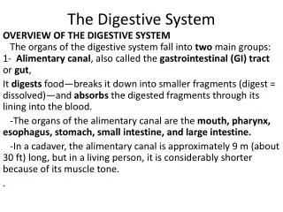
Chapter 22 – The Digestive System . 2 main components. Alimentary canal Continuous passageway from mouth to anus Accessory organs/structures Assists with the physical and/or chemical breakdown of food Teeth, salivary glands, pancreas, liver . Digestive processes. Ingestion

Chapter 22 – The Digestive System
E N D
Presentation Transcript
2 main components • Alimentary canal • Continuous passageway from mouth to anus • Accessory organs/structures • Assists with the physical and/or chemical breakdown of food • Teeth, salivary glands, pancreas, liver
Digestive processes • Ingestion • Bringing food into body’s interior • Digestion • Mechanical/physical • Breaking down into smaller components • Chewing/mastication • Stomach churning • Segmentation • Back and forth movement in small intestine • Chemical/enzymatic • Breaking of chemical bonds in nutrients into their smaller building blocks • Absorption • Nutrients pass through intestinal wall into bloodstream • Elimination • Removal of waste/undigested material • Defecation
Histology of alimentary canal • 4 tunics • Mucosa – deepest • Secretes mucus, enzymes • Contains lymphoid cells to fight infection • Submucosa • Blood and lymphatic vessels, nerve fibers • Muscularisexterna – smooth muscle • Deep – circular layer • Superficial – longitudinal layer • Serosa – most superficial • Visceral peritoneum • Reduces friction from organ movement
Peristalsis • Waves of smooth muscle contraction that propels the contents through the alimentary canal/GI tract
Mouth • Roof • Anterior hard palate • Maxillary and palatine bones • Posterior soft palate • Underlying muscle only; no bone • Uvula • Elicits gag response • Blocks entrance into nasopharynx when swallowing • Tongue • Moves food around during mastication to form bolus, and to push bolus to back of throat • Bolus – chewed food mixed with saliva • Deglutition = swallowing • Covered with taste buds • Also scattered in walls or oral cavity and oropharynx
Salivary glands • Produce saliva • Moistens food • Contains lysozyme to kill bacteria • Dissolves chemicals for sense of taste • Contains enzyme amylase • Begins chemical digestion of carbohydrates • 3 • Parotid • Sublingual • Submandibular
Tooth structure • Crown – exposed region; root – below gumline; neck – separates crown and root • Enamel covers crown • Calcium salts • Cementum – calcified connective tissue • Attachment site of periodontal ligament • Dentin – bulk of tooth • Similar to bone composition • Central pulp cavity • Extends through root canal • Vessels, nerve endings, connective tissue
Teeth #/type • Baby/ deciduous teeth • 20 total • 2-1-0-2 2-1-0-2 • Adult teeth • 32 total • 2-1-2-3 2-1-2-3
Pharynx and Esophagus Stratified squamous epithelium Esophageal/stomach junction • Pharynx • Oropharynx and laryngopharynx • Esophagus • Joins with stomach and cardiac orifice • Cardiac or gastroesophageal sphincter • Diaphragm helps keep it closed
Stomach • Muscularisexterna has a third layer – oblique (deep to circular layer) • Rugae – inner lining folds • Increases surface area • Allows stomach to expand • Regions • Cardia/cardiac • Fundus • Body • Lesser curvature – medial • Greater curvature – lateral • Pylorus • Plyoric sphincter at junction with small intestine • Food and gastric juice mix to form thick acid chyme • Exits from stomach in spurts • Higher fat content stays in stomach longer
Stomach histology • Mucous cells • Parietal cells • Secrete hydrochloric acid • Chief cells • Secrete pepsinogen • In presence of HCl, converts to active pepsin • Chemical digests protein
Small intestine • Site where most of chemical digestion occurs, and absorption of nutrients • 3 regions • Duodenum • Smallest section; site of most digestion • Entrance for hepatic and pancreatic secretions • Jejunum • Ileum • Contains Peyer’s patches • Large aggregates of lymphoid material • Ileocecal valve – junction with large intestine
Small intestine modifications for absorption • Microvilli – plasma membrane extensions of cell • Villi – projections of mucosa • Both villi and microvilli increase surface area • Pilcaecirculares • Allows chyme to swirl through lumen • Slows movement and increases contact with walls for absorption
Pancreas • Tail – next to spleen; body – surrounded by duodenum • Pancreatic secretions into duodenum • Bicarbonate ions • Neutralized acidity of chyme • Enzymes for digestion • Proteases, amylase, lipase
Liver • Main digestive function is to filter and process nutrient-rich blood from hepatic portal vein • Produces bile salts • Emulsifies fat • Mechanical breakdown of lipids into smaller droplets • Provides a greater surface area for enzymes to work on
Gallbladder • Concentrates and stores bile • Bile exits through cystic duct to bile duct, then into duodenum • Excess cholesterol can crystallize to form gallstones
Histology Pancreas Liver
Large intestine • Absorbs water and compacts waste • Contains bacteria • Produces B12 and K vitamins, which are then absorbed • Breakdown of molecules cause gaseous byproducts – flatus • Longitudinal layer of muscularis is reduced to three bands – teniae coli • Causes walls to ‘pucker’ into pouches called haustra • Epiploic appendages
Large intestine sections • Cecum “blind pouch” • Ileocecal valve • Vermiform appendix – mass of lymphoid tissue • Ascending colon – right side of body • Turns at hepatic flexure • Transverse colon • Turns at splenic flexure • Descending colon – left side of body • Sigmoid colon – “S” shape curves posteriorly
Large intestine sections cont • Rectum • Anus • Internal sphincter • Smooth muscle • External sphincter • Skeletal muscle
Chemical digestion • Large macromolecules (polymers) are broken down into monomer building blocks • Hydrolysis – addition of water molecule breaks one bond • Carbohydrates • Enzymatic breakdown begins in mouth with amylase • Monomer = monosaccharide • Glucose, fructose, galactose • Dissaccharide = 2 monosaccharides • Sucrose, lactose • Polysaccharide = complex carbohydrate • Glycogen, starch, cellulose (indigestible)
Chemical digestion cont • Proteins • Enzymatic breakdown begins in stomach by pepsin • monomer = amino acids • Lipids • Enzymatic breakdown begins in duodenum by lipase in adults • In children, begins in stomach • Monomer = fatty acid (and glycerol) • Nucleic acids • Enzymatic breakdown begins in duodenum • Does not contribute to cellular fuel/creation of ATP • Monomer = nucleotide