SKELETAL SYSTEM
Learn about the functions and organization of the skeletal system, including support and protection, body movement, blood cell formation, and storage of inorganic materials. Explore the axial and appendicular skeleton, types of bone tissue, bone development, and joint types.
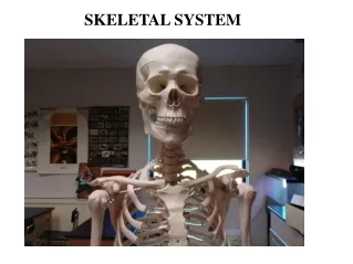

SKELETAL SYSTEM
E N D
Presentation Transcript
Functions of the Skeletal System Bones are made of OSSEOUS TISSUE Support and Protection Body movement Blood cell formation (bone marrow) - hematopoeisis Storage of inorganic materials (salt, calcium, potassium….)
ORGANIZATION About 206 bones 2 Main Divisions – Axial & Appendicular
Axial Skeleton Head, neck, trunk Skull Hyoid Bone Vertebral Column Thoracic Cage (ribs, 12 pairs) Sternum
Limbs & Bones that connect to the • Pectoral Girdle (shoulders) • Pelvic Girdle (hips) Appendicular Skeleton
Compact (wall of the diaphysis) Spongy (cancellous, epiphysis) - red marrow Types of Bone Tissue
Bone Details • Matrix: Where bone cells live • Osteocytes: Mature bone cells • All bones start as dense membranes • Primary Ossifications: shafts ( diaphysis) • Secondary Ossifications: ends (epiphesis)
Medullary Cavity – hollow chamber filled with bone marrow Red Marrow (blood) Yellow Marrow (fat) Endosteum – lining of the medullary Inside the Long Bone
Epiphysis: end part of long bone Diaphysis :Shaft of long bone Articular Cartilage: covers the articular surfaces of the bones participating in a synovial joint. Periosteum: dense layer of vascular connective tissue enveloping the bones except at the surfaces of the joints Endosteum layer of vascular tissue lining the inside of some bones ( also called medullary membrane) BONE STRUCTURE - Long Bone
EPIPHYSEAL DISK (growth plate) is a band of cartilage between the epiphysis (the end part of a long bone and the diaphysis (the shaft or central part of a long bone) These areas increase bone length as the cells ossify Cartilage becomes OSTEOBLASTS make new bone Osteoclasts: breaks down bones in reabsorption Bone Development & Growth
Synarthrotic (not moveable, aka sutures) • Amphiarthrotic (slightly moveable, vertebrae) • Diarthrotic (moveable joint, aka synovial joints) Types of Joints (articulations)
Synovial fluid - fluid within the joints that helps to lubricate Types of Joints 1. Ball and Socket 2. Hinge 3. Pivot 4. Saddle
BONES OF THE SKULL • Frontal: Anterior portion of eye2. Parietal – one on each side of skull3. Occipital –forms back of skull4. Temporal – forms sides and base of cranium5. Sphenoid – wedged between several bones6. Maxilla – forms upper jaw7. Mandible – forms lower jaw ( only moveable bone
Suture - refers to any connection between large bones (in fetal skulls, these are called fontanels) Fissure - any wide gap between bones
Sutures 1. Coronal - between frontal and parietal bones2. Lambdoidal - between occipital and parietal bones3. Squamosal - between temporal and parietal bones4. Sagittal - between parietal bones
Fontanels Cranium not solid at birth • Spaces called fontanels or “soft spots • Fibrous Tissue: allows for cranial expansion during birth ForanumMagnum Large opening where Spinal cord enters skull
Thoracic Cage • Thoracic Cage: 12 pairs of ribs • True ribs: first seven pairs attach directly to the sternum by vertebral sternal ( costal cartilage) • False ribs: last five pairs. Indirectly attached • Floating ribs : last two pairs within false ribs
Vertebrae Neck = cervical Middle Back = thoracic Lower Back = lumbar
Pectoral girdle • Shoulders • Two clavicles ( collar bones) • Two Scapulas ( shoulder blades) • Arms: humerus, radius and ulna • Wrist: 8 small bones called carpals • Fingers: phlanges
Ulna goes to pinky (P-U) Radius goes to thumb Bones of the Arm
For test Carpels Metacarpals Phalanges *extra credit opportunity Wrist Bones
Pelvic Girdle • Hips : Two large bones called coxal bones • Legs: Femur ( thigh bone), tibia & fibula ( lower leg • Ankle & upper foot :7 bones called tarsals • Largest is heel bone called the calcaneus, metatarsals and phlanges
Types of bone • Long Bones : Femur, Humerus and Tibia but are also some of the smallest includin the Metacarpals, Metatarsals and Phalanges. • Classification of a long bone includes having a body which is longer than it is wide, with growth plates (epiphysis) at either end.
Flat bones • Flat bones are as they sound, strong, flat plates of bone with the main function of providing protection to the bodies vital organs and being a base for muscular attachment. • The classic example of a flat bone is the Scapula (shoulder blade). The Sternum (breast bone), Cranium (skull), os coxae (hip bone) Pelvis and Ribs are also classified as flat bones. • Flat bones are as they sound, strong, flat plates of bone with the main function of providing protection to the bodies vital organs and being a base for muscular attachment. • The classic example of a flat bone is the Scapula (shoulder blade). The Sternum (breast bone), Cranium (skull), os coxae (hip bone) Pelvis and Ribs are also classified as flat bones. • Flat bones are as they sound, strong, flat plates of bone with the main function of providing protection to the bodies vital organs and being a base for muscular attachment. • The classic example of a flat bone is the Scapula (shoulder blade). The Sternum (breast bone), Cranium (skull), os coxae (hip bone) Pelvis and Ribs are also classified as flat bones.
Irregular Bones • These are bones in the body which do not fall into any other category, due to their non-uniform shape. Good examples of these are the Vertebrae, Sacrum and Mandible (lower jaw). They primarily consist of cancellous bone, with a thin outer layer of compact bone.
Sesamoid Bones • Sesamoid bones are usually short or irregular bones, imbedded in a tendon. The most obvious example of this is the Patella (knee cap) which sits within the Patella or Quadriceps tendon.
Abnormal Bone Conditions BONE SPURS: abnormal growth. Can occur on any bone (e.g. heel). OSTEOPOROSIS: Increased activity of osteoclasts cause a break down bone, and the subsequent fewer minerals in the extracellular matrix make it fragile. The spongy bone especially becomes more porous. Men get it as well as women. What’s the best way to prevent osteoporosis? Exercise! What does exercise do? Makes bones bigger. The most common bone used for a bone graft is the iliac bone of the hip.
Osteoporosis Figure 6.15
Rheumatoid arthritis is an autoimmune disease which causes joint stiffness and bone deformity Source: http://www.thetimes.co.uk/tto/public/article3233439.ece
ABNORMALITIES OF THE SPINE ABNORMALITIES OF THE SPINE SCOLIOSIS is a lateral curve in the spine KYPHOSIS is a hunchback curve LORDOSIS is a swayback in the lower region. ANKYLOSIS is severe arthritis in the spine and the vertebrae fuse.
FUN FACTS ABOUT BONESBone is made of the same type of minerals as limestone. Babies are born with 300 bones, but by adulthood we have only 206 in our bodies. The giraffe has the same number of bones in its neck as a human: seven in total. The long horned ram can take a head butt at 25 mph. The human skull will fracture at 5mph.


