Download
1 / 58
740 likes | 1.59k Vues
Childhood Atopic Dermatitis. Medical Student Core Curriculum In Dermatology. Last updated July 29, 2011. Module Instructions.

E N D
Childhood Atopic Dermatitis Medical Student Core Curriculum In Dermatology Last updated July 29, 2011
Module Instructions • The following module contains a number of blue, underlined terms which are hyperlinked to the dermatology glossary, an illustrated interactive guide to clinical dermatology and dermatopathology. • We encourage the learner to read all the hyperlinked information.
Goals and Objectives • The purpose of this module is to help medical students develop a clinical approach to the evaluation and initial management of patients presenting with atopic dermatitis. • After completing this module, the learner will be able to: • Identify and describe the morphology of atopic dermatitis • Recognize that superficial infections often complicate atopic dermatitis. • Recommend an initial treatment plan for a child with atopic dermatitis • Provide patient/parent education about daily skin care for a child with atopic dermatitis • Determine when to refer to a patient with atopic dermatitis to a dermatologist
Case One Carolyn Ku
Case One: History • HPI: Carolyn is a 10-month-old girl who was brought to the pediatric clinic by her mother for an “itchy red rash” for the last 7 months. The rash waxes and wanes, involving Carolyn’s face. Her mother reports Carolyn is bathed daily using a “normal” soap. Sometimes they use moisturizing lotion if her skin appears dry. They recently introduced peas into her diet and wonder whether this may be contributing to the rash. • PMH: Normal birth history. She is healthy aside from an episode of wheezing at 5 months of age. No hospitalizations or surgeries. • Medications: none • Allergies: none • Family history: Mother has asthma and allergic rhinitis • Social history: Lives in a house with her parents, no pets or recent travel • ROS: “itches all night”
Case One: Skin Exam How would you describe her skin exam?
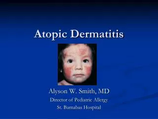
Case One: Skin Exam Erythematous ill-defined plaques with overlying scale and crust on her cheeks
Case One, Question 1 • What elements in the history are important to ask in this case? • Does she scratch or rub her skin? • Does the rash keep her awake at night? • Which moisturizers are used and where? • All of the above
Case One, Question 1 Answer: d • What elements in the history are important to ask in this case? • Does she scratch or rub her skin? (Provides information about associated pruritus, which will impact treatment) • Does the rash keep her awake at night? (Provides information about severity, which will impact treatment) • Which moisturizers are used and where? (May provide information about the distribution. Also, lack of using a moisturizer may be exacerbating the problem) • All of the above
Case One, Question 2 • What is the most likely diagnosis given the history and skin exam findings? • Atopic dermatitis • Contact dermatitis • Psoriasis • Scabies • Seborrheic dermatitis
Case One, Question 2 Answer: a • What is the most likely diagnosis given the history and skin exam findings? • Atopic dermatitis • Contact dermatitis(would expect history of contact with allergen and erythema with superimposed vesicles or bullae) • Psoriasis(presents as erythematous plaques with an adherent silvery scale) • Scabies(intensely pruritic papules, often with excoriation, burrows may be present) • Seborrheic dermatitis(would expect erythematous patches and plaques with greasy, yellowish scale)
Case One, Question 3 • Which of the following statements supports the diagnosis of atopic dermatitis: • Chronic nature of the rash • Distribution of the rash • Family history of atopic disease • Symptom of pruritus • All of the above
Case One, Question 3 Answer: e • Which of the following statements supports the diagnosis of atopic dermatitis: • Chronic nature of the rash (present x 7 months) • Distribution of the rash (predominantly on the cheeks) • Family history of atopic disease • Symptom of pruritus (itching) • All of the above
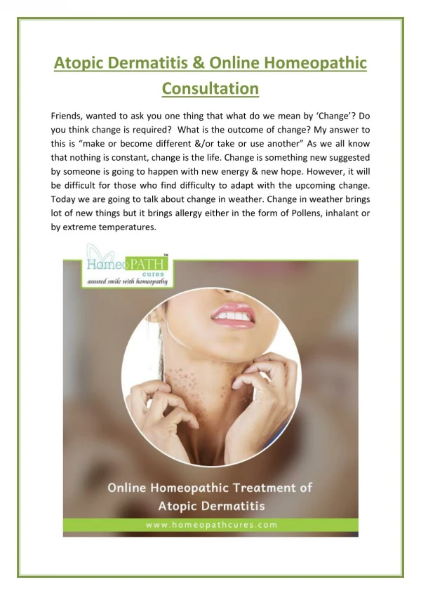
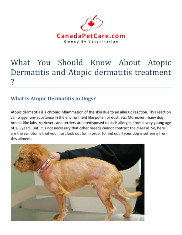
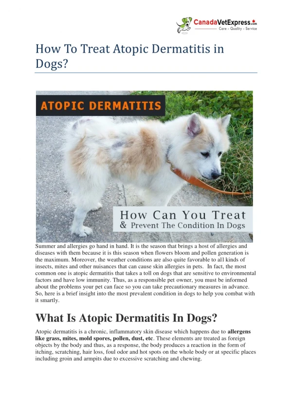
Atopic Dermatitis: The Basics • Atopic dermatitis (AD) is a chronic, pruritic, inflammatory skin disease with a wide range of severity • AD is one of the most common skin disorders in developed countries, affecting up to 20% of children & 1-3 % of adults • In most patients, AD develops before the age of 5 and typically clears by adolescence • Primary symptom is pruritus (itch) • AD is often called “the itch that rashes” • Scratching to relieve AD-associated itch gives rise to the ‘itch-scratch’ cycle and can exacerbate the disease • Patients experience periods of remission and exacerbation
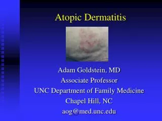
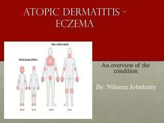
AD: Clinical Findings • Lesions typically begin as erythematous papules, which then coalesce to form erythematous plaques that may display weeping, crusting, or scale • Distribution of involvement varies by age: • Infants and toddlers: eczematous plaques appear on the cheeks forehead, scalp and extensor surfaces • Older children and adolescents: lichenified, eczematous plaques in flexural areas of the neck, elbows, wrists, and ankles • Adults: lichenification in flexural regions and involvement of the hands, wrists, ankles, feet, and face (particularly the forehead and around the eyes) • Xerosis is a common characteristic of all stages
Case One, Question 4 • What percentage of children with atopic dermatitis also have or will develop asthma or allergic rhinitis? • 0-15% • 15-30% • 30-50% • 50-80% • 80-100%
Case One, Question 4 The Atopic Triad Asthma Atopic dermatitis Allergic rhinitis Answer: d • What percentage of children with atopic dermatitis also have or will develop asthma or allergic rhinitis? • 50-80% of children will have another atopic disease
Typical AD for Infants and Toddlers Affects the cheeks, forehead, scalp, and extensor surfaces Erythematous, ill-defined plaques on the cheeks with overlying scale and crusting Erythematous, ill-defined plaques on the lateral lower leg with overlying scale
More Examples of Atopic Dermatitis Note the distribution of face and extensor surfaces
Typical AD for Older Children Affects flexural areas of neck, elbows, knees, wrists, and ankles Lichenified, erythematous plaques behind the knees Erythematous, excoriated papules with overlying crust in the antecubital fossa
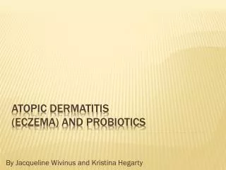
Atopic Dermatitis ≠ Eczema • Eczema is a nonspecific term that refers to a group of inflammatory skin conditions characterized by pruritus, erythema, and scale. • Atopic dermatitis is a specific type of eczematous dermatitis.
Atopic Dermatitis: Pathogenesis • The cause of AD is multifactorial and not completely understood • The following factors are thought to play varying roles: • Genetics • Skin Barrier Dysfunction • Impaired Immune Response • Environment
Back to Case One Carolyn Ku
Case One, Question 5 • Which of the following recommendations would you provide to Carolyn’s parents? • Daily or twice daily application of moisturizing ointment or cream • Hydrocortisone 2.5% ointment to the face twice daily • Hydroxyzine 1 tsp. (1mg/kg) PO at bedtime • Mild cleanser, as little as needed to remove dirt • All of the above
Case One, Question 5 Answer: e • Which of the following recommendations would you provide to Carolyn’s parents? • Daily or twice daily application of moisturizing ointment or cream • Hydrocortisone 2.5% ointment to the face twice daily • Hydroxyzine 1 tsp. (1mg/kg) PO at bedtime • Mild cleanser, as little as needed to remove dirt • All of the above • Carolyn is having an exacerbation of her AD and needs both gentle skin care and treatment of the inflammation in her skin
Atopic Dermatitis: Treatment • Combination of short-term treatment to manage flares and longer-term strategies to help control symptoms between flares • Recommend gentle skin care • Tepid baths without washcloths or brushes • Mild soaps • Pat dry • Emollients: petrolatum and moisturizers • Use ointments or thick creams (no watery lotions) • Apply once to twice daily to whole body (within 3 minutes of bathing for optimal occlusion) • Identification and avoidance of triggers and irritants (such as wool and acrylic fabrics)
AD: Treatment Continued • Treat acute inflammation with topical corticosteroids • Ointments are preferred over creams • Low potency is usually effective for the face • Body and extremities often require medium potency • Using stronger steroid for short periods and milder steroid for maintenance helps reduce risk of steroid atrophy and other side effects • Potential local side effects associated with topical corticosteroid therapy use include striae, telangiectasias, atrophy, and acne • Topical calcineurin inhibitors: 2nd-line therapy • Use when the continued use of topical steroids is ineffective or when the use of topical steroids is inadvisable
AD: Treatment Continued • Treat pruritus with antihistamines • Antihistamines help to break the itch/scratch cycle • Standing night-time 1st generation H1 antihistamines (e.g. hydroxyzine) are helpful • Treat co-existing skin infection with systemic antibiotics • Patients should be referred to a dermatologist when: • Patients have recurrent skin infections • Patients have extensive and/or severe disease • Symptoms are poorly controlled with topical steroids
Case One, Question 5 • Clobetasol ointment • Fluocinonide ointment • Hydrocortisone cream • Hydrocortisone ointment • Triamcinolone ointment • What is the most likely corticosteroid you would choose for Carolyn’s facial lesions?
Case One, Question 5 • Clobetasol ointment • Fluocinonide ointment • Hydrocortisone cream • Hydrocortisone ointment • Triamcinolone ointment Answer: d • What is the most likely corticosteroid you would choose for Carolyn’s facial lesions?
Topical Steroid Strength • Remember to look at the class not the percentage • Note that clobetasol 0.05% is stronger than hydrocortisone 1% • When several are listed, they are listed in order of strength • Note that triamcinolone ointment is stronger than triamcinolone cream or lotion because of the nature of the vehicle
Case One, Question 6 • Which of the following prescriptions should be used to treat Carolyn’s AD for a 3 month duration? • Hydrocortisone 2.5% cream, apply to affected area BID, dispense 30 grams • Hydrocortisone 2.5% cream, apply to affected area BID, dispense 90 grams • Hydrocortisone 2.5% ointment, apply to affected area BID, dispense 30 grams • Hydrocortisone 2.5% ointment, apply to affected area BID, dispense 90 grams
Case One, Question 6 Answer: d • Which of the following prescriptions should be used to treat Carolyn’s AD for a 3 month duration? • Hydrocortisone 2.5% cream, apply to affected area BID, dispense 30 grams • Hydrocortisone 2.5% cream, apply to affected area BID, dispense 90 grams • Hydrocortisone 2.5% ointment, apply to affected area BID, dispense 30 grams • Hydrocortisone 2.5% ointment, apply to affected area BID, dispense 90 grams (~ 2% BSA = 30 grams for 1 month x3 = 90 grams. Ointment is more occlusive) Refer to the Dermatologic Therapies module for more information about calculating amounts of topical medication
Topical Steroid Dosing in Children • Low potency topical corticosteroids are safe when used for short intervals • Can cause side effects when used for extended durations • High potency steroids must be used with caution and vigilant clinical monitoring for side effects in children • Potent steroids should be avoided in high risk areas such as the face, folds, or occluded areas such as under the diaper
Parent education and written instruction are key to success • “Action Plans” provide parents and caregivers with easy to follow treatment recommendations and guidance
Case One, Question 7 • Carolyn’s parents would also like more information regarding the association between food allergies and atopic dermatitis. What can you tell them? • A positive allergen test proves that the allergy is clinically relevant • Elimination of food allergens in patients with AD and confirmed food allergy will not lead to clinical improvement • Food allergy is a more likely trigger if the onset or worsening of the AD correlates with exposure to the food • There is no correlation between AD and food allergies
Case One, Question 7 Answer: c • Carolyn’s parents would also like more information regarding the association between food allergies and atopic dermatitis. What can you tell them? • A positive allergen test proves that the allergy is clinically relevant (Not true) • Elimination of food allergens in patients with AD and confirmed food allergy will not lead to clinical improvement (Not true. If the food allergy is clinically relevant, then the elimination of the food allergen will lead to improvement) • Food allergy is a more likely trigger if the onset or worsening of the AD correlates with exposure to the food • There is no correlation between AD and food allergies (Not true)
Allergens and Atopic Dermatitis • The role of allergy in AD remains controversial • Many patients with AD have sensitization to food and environmental allergens • However, evidence of allergen sensitization is not proof of a clinically relevant allergy • Food allergy as a cause of, or exacerbating factor for, AD is uncommon • Identification of true food allergies should be reserved for refractory AD in children in whom the suspicion for a food allergy is high • Infants with AD and food allergy may have additional findings that suggest the presence of food allergy, such as vomiting, diarrhea, and failure to thrive • Elimination of food allergens in patients with AD and confirmed food allergycan lead to clinical improvement
Case Two Joanna Shafer
Case Two: History • HPI: Joanna Shafer is a 10-year-old girl with a history of atopic dermatitis, normally well-controlled with emollients and occasional topical steroids who was brought in by her mother with an itchy red rash on the back of her thighs. • PMH: atopic dermatitis • Medications: hydrocortisone 2.5% ointment • Allergies: none • Family history: little sister with atopic dermatitis • Social history: Lives in a house with parents and sister. Attends fourth grade, favorite subject in school is spelling. • ROS: no fevers
Case Two: Skin Exam Multiple erythematous papules and plaques with erosions
Case Two, Question 1 • What is your next step in the evaluation of Joanna’s skin condition? • Apply a potent topical corticosteroid • Obtain a skin bacterial culture • Skin biopsy • Start topical antibiotics • None of the above
Case Two, Question 1 Answer: b • What is your next step in the evaluation of Joanna’s skin condition? • Apply a potent topical corticosteroid (will not help with evaluation) • Obtain a skin bacterial culture • Skin biopsy (not necessary for diagnosis) • Start topical antibiotics (a large majority of patients with AD are colonized with S. aureus, treating locally with topical antibiotics is usually not effective) • None of the above
Case Two: Evaluation • Skin bacterial culture should be considered during hyperacute, weepy flares of AD and when pustules or extensive yellow crust are present • Patients with AD are susceptible to a variety of secondary cutaneous infections such as Staphylococcus aureus and Group A Streptococcal infections • These infections are a common cause of AD exacerbations • Systemic antibiotics should be used to treat these infections
Case Three Mark Maldonado
Case Three: History • HPI: Mark is a 9-year-old boy who was brought in by his father who is concerned about the “white spots” on Mark’s face • PMH: mild asthma, no history of hospitalizations • Medications: albuterol when needed • Allergies: none • Family history: mother had a history of childhood atopic dermatitis • Social history: lives at home with his mother and father • ROS: negative
Case Three, Question 1 How would you describe Mark’s skin exam?
Case Three: Skin Exam Poorly defined hypopigmented, scaly patches on the face