Download
1 / 52
550 likes | 1.15k Vues
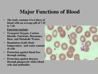
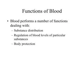
Functions of Blood. Blood performs a number of functions dealing with: Substance distribution Regulation of blood levels of particular substances Body protection. Blood Functions: Distribution. Blood transports: Oxygen from the lungs and nutrients from the digestive tract

E N D
Functions of Blood • Blood performs a number of functions dealing with: • Substance distribution • Regulation of blood levels of particular substances • Body protection
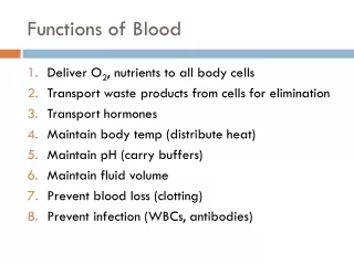
Blood Functions: Distribution • Blood transports: • Oxygen from the lungs and nutrients from the digestive tract • Metabolic wastes from cells to the lungs and kidneys for elimination • Hormones from endocrine glands to target organs
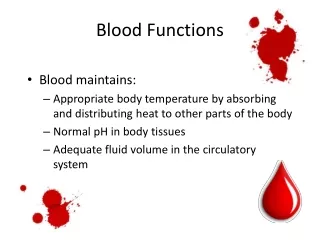
Blood Functions: Regulation • Blood maintains: • Appropriate body temperature by absorbing and distributing heat to other parts of the body • Normal pH in body tissues using buffer systems • Adequate fluid volume in the circulatory system
Blood Functions: Protection • Blood prevents blood loss by: • Activating plasma proteins and platelets • Initiating clot formation when a vessel is broken • Blood prevents infection by: • Synthesizing and utilizing antibodies • Activating complement proteins • Activating WBCs to defend the body against foreign invaders
Physical Characteristics of Blood • Average volume of blood: • 5–6 L for males; 4–5 L for females (Normovolemia) • Hypovolemia - low blood volume • Hypervolemia - high blood volume • Viscosity (thickness) - 4 - 5 (where water = 1) • The pH of blood is 7.35–7.45; x = 7.4 • Osmolarity = 300 mOsm or 0.3 Osm • This value reflects the concentration of solutes in the plasma • Salinity = 0.85% • Reflects the concentration of NaCl in the blood • Temperature is 38C, slightly higher than “normal” body temperature • Blood accounts for approximately 8% of body weight
Composition of Blood • Blood is the body’s only fluid tissue (a connective tissue) • 2 major components • Liquid = plasma (55%) • Formed elements (45%) • Erythrocytes, or red blood cells (RBCs) • Leukocytes, or white blood cells (WBCs) • Platelets - fragments of megakaryocytes in marrow
Components of Whole Blood Plasma(55% of whole blood) Buffy coat:leukocyctes and platelets(<1% of whole blood) Formed elements Erythrocytes(45% of whole blood) Withdraw blood and place in tube Centrifuge 1 2 • Hematocrit • Males: 47% ± 5% • Females: 42% ± 5%
Blood Plasma • Blood plasma components: • Water = 90-92% • Proteins = 6-8% • Albumins; maintain osmotic pressure of the blood • Globulins • Alpha and beta globulins are used for transport purposes • Gamma globulins are the immunoglobulins (IgG, IgA, etc) • Fibrinogen; a clotting protein • Organic nutrients – glucose, carbohydrates, amino acids • Electrolytes – sodium, potassium, calcium, chloride, bicarbonate • Nonprotein nitrogenous substances – lactic acid, urea, creatinine • Respiratory gases – oxygen and carbon dioxide
Formed Elements • Formed elements comprise 45% of blood • Erythrocytes, leukocytes, and platelets make up the formed elements • Only WBCs are complete cells • RBCs have no nuclei or organelles, and platelets are just cell fragments • Most formed elements survive in the bloodstream for only a few days • Most blood cells do not divide but are renewed by cells in bone marrow
Erythrocytes (RBCs) • Biconcave disc • Folding increases surface area (30% more surface area) • Plasma membrane contains spectrin • Give erythrocytes their flexibility • Anucleate, no centrioles, no organelles • End result - no cell division • No mitochondria means they generate ATP anaerobically • Prevents consumption of O2 being transported • Filled with hemoglobin (Hb) - 97% of cell contents • Hb functions in gas transport • Hb + O2 HbO2 (oxyhemoglobin) • Most numerous of the formed elements • Females: 4.3–5.2 million cells/cubic millimeter • Males: 5.2–5.8 million cells/cubic millimeter
Erythrocytes (RBCs) Figure 17.3
Erythrocyte Function • Erythrocytes are dedicated to respiratory gas transport • Hemoglobin reversibly binds with oxygen and most oxygen in the blood is bound to hemoglobin • Composition of hemoglobin • A protein called globin • made up of two alpha and two beta chains • A heme molecule • Each heme group bears an atom of iron, which can bind to one oxygenmolecule • Each hemoglobin molecule thus can transport four molecules of oxygen
Structure of Hemoglobin Figure 17.4
Hemoglobin • Oxyhemoglobin – hemoglobin bound to oxygen • Oxygen loading takes place in the lungs • Deoxyhemoglobin – hemoglobin after oxygen diffuses into tissues (reduced Hb) • Carbaminohemoglobin – hemoglobin bound to carbon dioxide • Carbon dioxide loading takes place in the tissues
Fate and Destruction of Erythrocytes • The life span of an erythrocyte is 100–120 days • Travels about 750 miles in that time (LA to Albuquerque) • Old erythrocytes become rigid and fragile, and their hemoglobin begins to degenerate • Dying erythrocytes are engulfed by macrophages • Heme and globin are separated • Iron is removed from the heme and salvaged for reuse • Stored as hemosiderin or ferritin in tissues • Transported in plasma by beta-globulins as transferrin
Fate and Destruction of Erythrocytes • Heme is degraded to a yellow pigment called bilirubin • Liver secretes bilirubin into the intestines as bile • Intestines metabolize bilirubin into urobilinogen • Urobilinogen leaves the body in feces, in a pigment called stercobilin • Globin is metabolized into amino acids which are then released into the circulation
Stages of Differentiation of Blood Cells Figure 17.9
Production of Erythrocytes • Hematopoiesis – blood cell formation • Occurs in the red bone marrow (myeloid tissue) • Axial skeleton and girdles • Epiphyses of the humerus and femur • Marrow contains immature erythrocytes • Composed of reticular connective tissue • Hemocytoblasts give rise to ALL formed elements • Lymphoid stem cells - give rise to lymphocytes • Myeloid stem cells - give rise to all other blood cells
Production of Erythrocytes: Erythropoiesis • A hemocytoblast is transformed into a committed cell called the proerythroblast • Proerythroblasts develop into early erythroblasts • The developmental pathway consists of three phases • Phase 1 – ribosome synthesis in early erythroblasts • Phase 2 – hemoglobin accumulation in late erythroblasts and normoblasts • Phase 3 – ejection of the nucleus from normoblasts and formation of reticulocytes • Reticulocytes then become mature erythrocytes • Reticulocytes make up about 1 -2 % of all circulating erythrocytes
Regulation and Requirements for Erythropoiesis • Circulating erythrocytes – the number remains constant and reflects a balance between RBC production and destruction • Too few red blood cells leads to tissue hypoxia • Too many red blood cells causes undesirable blood viscosity • Erythropoiesis is hormonally controlled and depends on adequate supplies of iron, amino acids, and B vitamins
Hormonal Control of Erythropoiesis • Erythropoietin (EPO) release by the kidneys is triggered by: • Hypoxia due to decreased RBCs • Decreased oxygen availability • Increased tissue demand for oxygen • Enhanced erythropoiesis increases the: • RBC count in circulating blood • Oxygen carrying ability of the blood
Erythropoietin Mechanism Imbalance Start Normal blood oxygen levels Stimulus: Hypoxia due to decreased RBC count, decreased availability of O2 to blood, or increased tissue demands for O2 Imbalance Increases O2-carrying ability of blood Reduces O2 levels in blood Erythropoietin stimulates red bone marrow Kidney (and liver to a smaller extent) releases erythropoietin Enhanced erythropoiesis increases RBC count Figure 17.6
Dietary Requirements of Erythropoiesis • Erythropoiesis requires: • Proteins, lipids, and carbohydrates • Iron, vitamin B12, and folic acid • The body stores iron in Hb (65%), the liver, spleen, and bone marrow • Intracellular iron is stored in protein-iron complexes such as ferritin and hemosiderin • Circulating iron is loosely bound to the transport protein transferrin
Erythrocyte Disorders • Polycythemia • Abnormal excess of erythrocytes • Increases viscosity, decreases flow rate of blood • Anemia – blood has abnormally low oxygen-carrying capacity • It is a symptom rather than a disease itself • Blood oxygen levels cannot support normal metabolism • Signs/symptoms include fatigue, paleness, shortness of breath, and chills
Anemia: Insufficient Erythrocytes • Hemorrhagic anemia – result of acute or chronic loss of blood • Hemolytic anemia – prematurely ruptured erythrocytes • Aplastic anemia – destruction or inhibition of red bone marrow
Anemia: Decreased Hemoglobin Content • Iron-deficiency anemia results from: • A secondary result of hemorrhagic anemia • Inadequate intake of iron-containing foods • Impaired iron absorption • Pernicious anemia results from: • Deficiency of vitamin B12 • Lack of intrinsic factor needed for absorption of B12 • Treatment is intramuscular injection of B12
Anemia: Abnormal Hemoglobin • Thalassemias – absent or faulty globin chain in hemoglobin • Erythrocytes are thin, delicate, and deficient in hemoglobin • Sickle-cell anemia – results from a defective gene • Codes for an abnormal hemoglobin called hemoglobinS (HbS) • This defect causes RBCs to become sickle-shaped in low oxygen situations
Polycythemia • Polycythemia – excess RBCs that increase blood viscosity • Three main polycythemias are: • Polycythemia vera • Secondary polycythemia • Blood doping
Leukocytes (WBCs) • Leukocytes, the only blood components that are complete cells: • 4,800 - 10,000/cubic millimeter • Protect the body from infectious microorganisms • Can leave capillaries via diapedesis • Move through tissue spaces (amoeboid motion) • Many are phagocytic (possess numerous lysosomes) • Two major types of leukocytes • Granulocytes: Neutrophils, Eosinophils, Basophils • Agranulocytes: Monocytes, Lymphyocytes • Leukocytosis – WBC count over 11,000/mm3 • Normal response to bacterial or viral invasion • Leukopenia - a decrease in WBC count below 4,800/mm3 • Leukemia - a cancer of WBC
Granulocytes • Granulocytes – neutrophils, eosinophils, and basophils • Contain cytoplasmic granules that stain specifically (acidic, basic, or both) with Wright’s stain • Are larger and usually shorter-lived than RBCs • Have lobed nuclei • Are all phagocytic cells
Granulocytes: Neutrophils(Polymorphonuclear leukocytes) • Account for 65-75% of total WBC’s • Neutrophils have two types of granules that: • Take up both acidic and basic dyes • Give the cytoplasm a lilac color • Contain peroxidases, hydrolytic enzymes, and defensins (antibiotic-like proteins) • Neutrophils are our body’s bacteria slayers • AKA “polys” or PMN’s (polymorphonuclear)
Granulocytes: Eosinophils • Eosinophils account for 1–4% of WBCs • Have red-staining, bilobed nuclei • Have red to crimson granules • Function: • Lead the body’s counterattack against parasitic infections • Lessen the severity of allergies by phagocytizing immune complexes (ending allergic reactions)
Granulocytes: Basophils • Account for 0.5-1% of all WBCs • Have U- or S-shaped nuclei with two or three conspicuous constrictions • Are functionally similar to mast cells • Have large, purplish-black (basophilic) granules that contain histamine • Histamine – inflammatory chemical that acts as a vasodilator and attracts other WBCs (antihistamines counter this effect)
Agranulocytes: Lymphocytes • Account for 20-25% or more of WBCs and: • Have large, dark-purple, circular nuclei with a thin rim of blue cytoplasm • Are found mostly enmeshed in lymphoid tissue (some circulate in the blood) • Most important cells of the immune system • There are two types of lymphocytes: T cells and B cells • T cells - attack foreign cells directly • B cells give rise to plasma cells, which produce antibodies
Monocytes • Monocytes account for 3–7% of leukocytes • They are the largest leukocytes • They have purple-staining, U- or kidney-shaped nuclei • They leave the circulation, enter tissue, and differentiate into macrophages
Production of Leukocytes • Leukopoiesis is hormonally stimulated by two families of cytokines (hematopoietic factors) – interleukins and colony-stimulating factors (CSFs) • Interleukins are numbered (e.g., IL-1, IL-2), whereas CSFs are named for the WBCs they stimulate (e.g., granulocyte-CSF stimulates granulocytes) • Macrophages and T cells are the most important sources of cytokines • Many hematopoietic hormones are used clinically to stimulate bone marrow
Formation of Leukocytes • All leukocytes originate from hemocytoblasts • The mother of all blood stem cells • Hemocytoblasts differentiate into myeloid stem cells and lymphoid stem cells • Myeloid stem cells become myeloblasts or monoblasts • Granulocytes form from myeloblasts • Monoblasts enlarge and form monocytes • Lymphoid stem cells become lymphoblasts • Lymphoblasts develop into lymphocytes
Formation of Leukocytes Figure 17.11
Leukocytes Disorders: Leukemias • Leukemia refers to cancerous conditions involving white blood cells • Leukemias are named according to the abnormal white blood cells involved • Myelocytic leukemia – involves myeloblasts • Lymphocytic leukemia – involves lymphocytes • Acute leukemia involves blast-type cells and primarily affects children • Chronic leukemia is more prevalent in older people
Leukemia • Immature white blood cells are found in the bloodstream in all leukemias • Bone marrow becomes totally occupied with cancerous leukocytes • Severe anemia ensues due to excess production of WBC’s • The white blood cells produced, though numerous, are not functional • Death is caused by internal hemorrhage and overwhelming infections • Treatments include irradiation, antileukemic drugs, and bone marrow transplants
Platelets • Platelets are fragments of megakaryocytes • Their granules contain serotonin, Ca2+, enzymes, ADP, and platelet-derived growth factor (PDGF) • Platelets function in the clotting mechanism by forming a temporary plug that helps seal breaks in blood vessels • Platelets not involved in clotting are kept inactive by Nitric Oxide (NO) and prostaglandins
Human Blood Groups • RBC membranes have glycoprotein antigens on their external surfaces • These antigens are: • Unique to the individual • Recognized as foreign if transfused into another individual • Promoters of agglutination and are referred to as agglutinogens • Presence or absence of these antigens is used to classify blood groups
Blood Groups • Humans have 30 varieties of naturally occurring RBC antigens • The antigens of the ABO and Rh blood groups cause vigorous transfusion reactions when they are improperly transfused • Other blood groups (M, N, Dufy, Kell, and Lewis) are mainly used for legalities
ABO Blood Groups • The ABO blood groups consists of: • Two antigens (A and B) on the surface of the RBCs • Two antibodies in the plasma (anti-A and anti-B) • An individual with ABO blood may have various types of antigens and spontaneously preformed antibodies • Agglutinogens and their corresponding antibodies cannot be mixed without serious hemolytic reactions
ABO Blood Groups Table 17.4
Rh Blood Groups • Presence of the Rh agglutinogens on RBCs is indicated as Rh+; 85% of population is + • Lack of antigen indicated as Rh -; 15% of popn. • Anti-Rh antibodies are not spontaneously formed only in Rh– individuals • However, if an Rh– individual receives Rh+ blood, anti-Rh antibodies form • A second exposure to Rh+ blood will result in a typical transfusion reaction
Hemolytic Disease of the Newborn • May occur in an Rh- mom pregnanet with an Rh+ fetus • Hemolytic disease of the newborn – Rh+ antibodies of a sensitized Rh– mother cross the placenta and attack and destroy the RBCs of an Rh+ baby • Rh– mother becomes sensitized when Rh+ blood (from a previous pregnancy of an Rh+ baby or a Rh+ transfusion) causes her body to synthesis Rh+ antibodies • The drug RhoGAM can prevent the Rh– mother from becoming sensitized • Treatment of hemolytic disease of the newborn involves pre-birth transfusions and exchange transfusions after birth
Transfusion Reactions • Transfusion reactions occur when mismatched blood is infused • Donor’s cells are attacked by the recipient’s plasma agglutinins causing: • Diminished oxygen-carrying capacity • Clumped cells that impede blood flow • Ruptured RBCs that release free hemoglobin into the bloodstream • Circulating hemoglobin precipitates in the kidneys and causes renal failure