Download

1 / 34
340 likes | 492 Vues
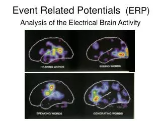
Event-Related Potentials (ERPs) are small electrical responses embedded within EEG signals, triggered by specific events such as stimulus presentations or motor actions. By averaging these responses across electrodes, we isolate ERP waveforms. While individual waveforms provide valuable data, they may not fully capture the overall potential patterns across the scalp. Techniques like Brain Electrical Source Analysis aim to reconstruct the 3D locations of electrical generators influencing these potentials. This analysis contributes significantly to our understanding of brain function in both human and animal studies.

E N D
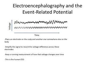
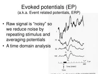
The Event-Related Potential (ERP) • Embedded in the EEG signal is the small electrical response due to specific events such as stimulus or task onsets, motor actions, etc.
The Event-Related Potential (ERP) • Embedded in the EEG signal is the small electrical response due to specific events such as stimulus or task onsets, motor actions, etc. • Averaging all such events together isolates this event-related potential
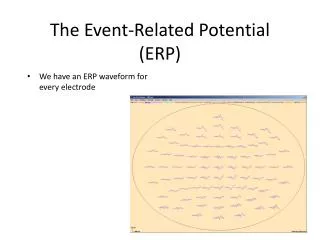
The Event-Related Potential (ERP) • We have an ERP waveform for every electrode
The Event-Related Potential (ERP) • We have an ERP waveform for every electrode
The Event-Related Potential (ERP) • We have an ERP waveform for every electrode • Sometimes that isn’t very useful
The Event-Related Potential (ERP) • We have an ERP waveform for every electrode • Sometimes that isn’t very useful • Sometimes we want to know the overall pattern of potentials across the head surface • isopotential map
The Event-Related Potential (ERP) • We have an ERP waveform for every electrode • Sometimes that isn’t very useful • Sometimes we want to know the overall pattern of potentials across the head surface • isopotential map Sometimes that isn’t very useful - we want to know the generator source in 3D
Brain Electrical Source Analysis • Given this pattern on the scalp, can you guess where the current generator was?
Brain Electrical Source Analysis • Given this pattern on the scalp, can you guess where the current generator was? • Source Imaging in EEG/MEG attempts to model the intracranial space and “back out” the configuration of electrical generators that gave rise to a particular pattern of EEG on the scalp Duracell
Brain Electrical Source Analysis • EEG data can be coregistered with high-resolution MRI image Source Imaging Result Structural MRI with EEG electrodes coregistered
Intracranial and “single” Unit • Single or multiple electrodes are inserted into the brain • “chronic” implant may be left in place for long periods
Intracranial and “single” Unit • Single electrodes may pick up action potentials from a single cell • An electrode may pick up thecombined activity from several nearby cells • spike-sorting attempts to isolate individual cells
Intracranial and “single” Unit • Simultaneous recording from many electrodes allows recording of multiple cells
Intracranial and “single” Unit • Output of unit recordings is often depicted as a “spike train” and measured in spikes/second • Spike rate is almost never zero, even without sensory input • in visual cortex this gives rise to “cortical grey” Stimulus on Spikes
Intracranial and “single” Unit • Local Field Potential reflects summed currents from many nearby cells Stimulus on Spikes
Relationship between EEG / LFP / spike trains • All three probably reflect related activities but probably don’t share a 1-to-1 mapping • For example: there could be some LFP or EEG signal that isn’t associated with a change in spike rates. • WHY? Whittingstall & Logothetis (2009)
Lesion Studies • Logic of Lesion Studies: • damaged area plays a role in accomplishing whatever task is deficient after the lesion
Lesion Studies • Types of Lesions • Animal • Human
Lesion Studies • Animal Lesion Techniques • Aspiration Lesions • Electrolytic Lesions
Lesion Studies • Animal Lesion Techniques • Aspiration Lesions • Electrolytic Lesions • Problems: • These can damage surrounding tissue - especially white matter tracts nearby (“fibers of passage”) • Irreversible • eventual degradation of connected areas
Lesion Studies • Animal Lesion Techniques • Vascular Lesions • endothelin-1 • good model of human stroke • severe damage • not pinpoint accuracy
Lesion Studies • Animal Lesion Techniques • Reversible Lesions • cooling • Local anesthetic, other drugs • highly selective • can cool specific layers of cortex • can be reversed!
Lesion Studies • Animal Lesion Techniques • Selective Pharmacological lesions • damage or destroy entire pathways that have a specific sensitivity to a particular chemical • e.g. MPTP model of Parkinson’s Disease (frozen addicts) • e.g. scapolomine - acetylcholine antagonist - temporary amnesia • Can be selective for specific circuits but not for specific brain areas • can be reversible in some cases (e.g. scopolamine, but not MPTP)
Lesion Studies • Animal Lesion Techniques • Gene Knock-Out/Knock-In (Transgenics) • can selectively block/enhance expression • Viral vectors, electroporation • animal develops differently • Can have temporal/regional/molecular specificity
Lesion Studies • Human Lesions • Ischemic Events • Stroke and Hemorrhage: • typically due to blood clot or hemorrhage • size of lesion depends on where clot gets lodged • amount of damage depends on how long clot remains lodged
Lesion Studies • Human Lesions • Trauma • Frontal lobes are particularly susceptible • Some famous cases (e.g. Phineas Gage)
Lesion Studies • Human Lesions • Surgery • Often surgery done to treat epilepsy • Occasionally corpus callosum is severed • Problem: patient wasn’t “normal” before the surgery
Lesion Studies • Human Lesions • Transcranial Magnetic Stimulation • Electromagnet Induces current in the brain • very transient, very focal reversible “lesion” • Believed to be safe • sites that can be studied are limited by the geometry of the head