Download
1 / 33
370 likes | 1.07k Vues
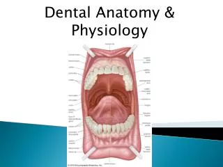
Human dentition Dental anatomy, physiology and occlusion. The Permanent Mandibular second Molars Dr. Samir M. Ziara B.D.S. (Alexandria Univ.) D. D. P. H. Royal Collage of Surgeon (London) M. Sc. P. H. Al-Quds Univ. Diploma of H. Administration. Mandibular Second Molar.

E N D
Human dentitionDental anatomy, physiology and occlusion The Permanent Mandibular second Molars • Dr. Samir M. Ziara • B.D.S. (Alexandria Univ.) • D. D. P. H. Royal Collage of Surgeon (London) • M. Sc. P. H. Al-Quds Univ. • Diploma of H. Administration
The mandibular second molar supplements the first molar in function. • Its anatomy differs in some details. • Normally, the second molar is smaller than the first molar by a fraction of a millimeter in all dimensions. • Although the roots are not as well formed, they may be longer.
The crown has four well-developed cusps: two buccal and two lingual, of nearly equal development. There is neither a distal nor a fifth cusp, but the distobuccal cusp is larger than that found on the first molar.The tooth has two well-developed roots, one mesial and one distal. These roots are broad buccolingually, but they are not as broad as those of the first molar, nor are they as widely separated.
Detailed Description of the Mandibular Second Molar from all Aspects In describing this tooth, direct comparisons will be made with the first mandibular molar.
Buccal Aspect • The crown is somewhat shorter cervico-occlusally and narrower mesiodistally than in the first molar. • The crown and root show a tendency toward greater overall length, but are not always longer. • There is but one developmental groove buccally, the buccal developmental groove. • This groove acts as a line of demarcation between the mesiobuccal and the distobuccal cusps, which are about equal in their mesiodistal measurements.
The roots may be shorter than those of the first molar, but they vary considerably in this as well as in their development generally. • The roots are usually closer together, and their axes are nearly parallel. • They may spread as much as those of the first molar or they may be fused for all or part of their length
The roots are inclined distally in relation to the occlusal plane of the crown, their axes forming more of an acute angle with the occlusal plane than is found on the first molar. • When one compares all of the mandibular molars, it may seem that the first molar shows one angulation of roots to occlusal plane. the second molar a more acute angle and the third molar an angle which is more acute still.
Lingual Aspect Differences in detail between the mandibular second molar and the mandibular first molar, to be noted from the lingual aspect. are these: • The crown and root of the mandibular second molar converge lingually but to a slight degree; little of the mesial or distal surfaces may therefore be seen from this aspect.
Lingual Aspect • The mesiodistal calibration at the cervix lingually is always greater accordingly than that of the first molar. • The curvatures mesially and distally on the crown that describe the contact areas are more noticeable from the lingual aspect. They prove to be at a slightly lower level, especially in the distal area, than those of the first molar
Mesial Aspect • Except for the differences in measurement from the mesial aspect, the second molar differs little from the first molar. • The cervical ridge buccally on the crown portion is in most instances less pronounced, and the occlusal surface may be more constricted buccolingually
Mesial Aspect • The cervical line shows less curvature, being straight and regular in outline buccolingually. • The mesial root is somewhat pointed apically. If part of the distal root is in sight, it is seen buccally. In the first molar, when the distal root is in sight from the mesial aspect, it is in view lingually.
Distal Aspect From the distal aspect, the second molar is similar in form to the first molar except for the • absence of a distal cusp and a distobuccal groove. • The contact area is centered on the distal surface buccolingually and is placed equidistant from cervical line and marginal ridge.
Occlusal Aspect • The occlusal aspect of the mandibular second molar differs considerably from the first molar. These variations serve as marks of identity. The small distal cusp of the first molar is not present, and the distobuccal lobe development is just as pronounced, and sometimes more so, than that of the mesiobuccal lobe.
Occlusal Aspect • There is no distobuccal developmental groove occlusally or buccally. • The buccal and lingual developmental grooves meet the central developmental groove at right angles at the central pit on the occlusal surface. • These grooves form a cross, dividing the occlusal portion of the crown into four parts
In general, the cusp slopes on the occlusal surface are not as smooth as those found on first molars, since they are roughened by many supplemental grooves radiating from the developmental grooves.
The following characteristics of mandibular second molars from the occlusal aspect should be observed and noted: • Many of them are rectangular from the occlusal aspect • Many show considerable prominence cervically on the mesiobuccal lobe only
Most second molars exhibit more curvature of the outline of the crown distally than mesially. showing a semicircular outline to the disto-occlusal surface in comparison with a square outline mesially. • The cusp ridge of the distobuccal cusp lies buccal to the cusp ridge of the mesiobuccal cusp
Human dentitionDental anatomy, physiology and occlusion The Permanent Mandibular third Molars • Dr. Samir M. Ziara • B.D.S. (Alexandria Univ.) • D. D. P. H. Royal Collage of Surgeon (London) • M. Sc. P. H. Al-Quds Univ. • Diploma of H. Administration
The Permanent Mandibular third Molars • The mandibular third molar varies considerably in different individuals and presents many anomalies both in form and in position. • It supplements the second molar in function, although the tooth is seldom as well developed, the average mandibular third molar showing irregular development of the crown portion, with undersized roots, more or less malformed.
Generally speaking, however, its design conforms to the general plan of all mandibular molars, conforming more closely to that of the second mandibular molar in the number of cusps and occlusal design than it does to the mandibular first molar. • Occasionally, mandibular third molars are seen that are well formed and comparable in size and development to the mandibular first molar
Many instances of mandibular third molars with five or more cusps are found, with the crown portions larger than those of the second molar. • In these cases, the alignment and occlusion with other teeth is not normal because insufficient room is available in the alveolar process of the mandible for the accommodation of such a large tooth and the occlusal form is too variable.
Although It is possible to find dwarfed specimens of mandibular third molars most of them that are not normal in size are larger than normal in the crown portion particularly. • Roots of these oversize third molars may be short and poorly formed. • The opposite situation is likely in maxillary third molars. Most of the anomalies are undersized.
Mandibular third molars are the most likely to be impacted, wholly or partially. • in the jaw. The lack of space accommodation is the chief cause.
If the third molar is congenitally absent from one side of the mandible or maxilla, it will most likely be absent from the other. • However. there is probably not a significant association between third molar agenesis in the maxilla and mandible.
Partial eruption of mandibular third molar teeth may result in periodontal defects on the distal aspects by the second molars, and in some instances resorption of distal root surfaces • When third molars are to be restored, it should be remembered that the depth of the enamel on the occlusal surface is relatively greater than first or second molars.
Detailed Description of the Mandibular Third Molar from All Aspects Buccal Aspect • From the buccal aspect, mandibular third molars vary considerably in outline. At the same time, they all have certain characteristics in common. • The outline of the crowns from this aspect is in a general way that of all mandibular molars.
The crown is wider at contact areas mesiodistally than at the cervix, the buccal cusps are short and rounded, and the crest of contour mesially and distally is located a little more than half the distance from cervical line to tips of cusps. • The type of third molar that is more likely to be in fair alignment and in good occlusion with other teeth is the four-cusp type; this is smaller and shows two buccal cusps only from this aspect
The average third molar also shows two roots, one mesial and one distal. • These roots are usually shorter, with a poorer development generally, than the roots of first or second molars, and their distal inclination in relation to the occlusal plane of the crown is greater. • The roots may be separated with a definite point of bifurcation, or they may be fused for all or part of their length
Lingual Aspect • Observations from the lingual aspect add little to those already made from the buccal aspect. • The mandibular third molar. when well developed. corresponds closely to the form of the second molar except for size and root development.
Mesial Aspect • From the mesial aspect. this tooth resembles the mandibular second molar except in dimensions. • The roots. of course. are shorter. with the mesial root tapering more from cervix to apex.' The apex of the mesial root is usually more pointed.
Distal Aspect • The anatomic appearance of the distal portion of this tooth is much like that of the second molar except for size. • Those specimens that have oversize crown portions are much more spheroidal above the cervical line. • The distal root appears smaIl. both in length and in buccolingual measurement when compared with the large crown portion.
Occlusal Aspect • The occlusal aspect is quite similar to that of the second mandibular molar • The tendency is toward a more rounded outline and a smaller buccolingual measurement distally.