Understanding the Compound Light Microscope: Magnification and Components
The compound light microscope is essential for studying small cells, as it uses two lenses to magnify objects, allowing for close examination. The ocular lens magnifies the image seen by the eye, while the objective lens, located just above the specimen, further magnifies it. Total magnification is calculated by multiplying the magnifications of both lenses. Key components include the stage for supporting slides, adjustment knobs for focusing, and the light source for illumination. This guide also covers practical exercises for enhancing your understanding of microscope usage.

Understanding the Compound Light Microscope: Magnification and Components
E N D
Presentation Transcript
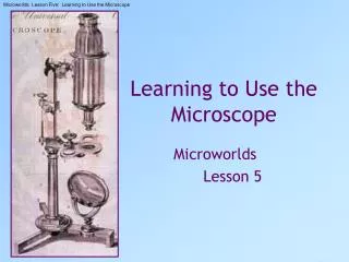
Because cells are small, you must make them appear larger than they really are in order to see and study them. To view cells closely, you will use a compound light microscope. It employs two lenses and a light source to make the object appear larger.
The object is magnified by a lens near your eye, the ocular lens (sometimes called the eyepiece), and again by a second lens, the objective lens, which is just above the object. The comparison of the actual size of the object with the size of its image is referred to as magnification.
Do you have difficulty imagining what 100X magnification looks like? The wing of a small butterfly, increased by 100X, would cover two cars. If it were increased to 1000X, it would cover four houses.
Two different lenses, the ocular and the objective lens, each magnify the object you are viewing. The total magnification of the image is determined by multiplying the two magnifications. For example, a magnification of 10X by the ocular lens and 4X by the objective lens would result in a magnification of the object of 40X (4 X 10).
Parts of the Microscope Ocular Lens – Magnifies the object. Also known as the eyepiece. Objective Lens – Magnify the object. Usually three complex lenses are located on the nosepiece immediately above the specimen. Stage – Supports the slide. A central opening in the stage allows light to pass through. Arm – Holds the ocular lens to the base and is the part that should be held firmly when moving the microscope from one location to another. Course-adjustment knob – Moves the tube up or down so you can get the specimen into focus. It is used with the low power objective lens only. Fine-adjustment knob –Moves the tube to get the object into sharp focus. It is used with medium- and high-power magnification. Use only after the specimen has been located and focused under low-power magnification. Condenser Lens – Directs light to the object. Light Source – Provides light for viewing the specimen. Power Cord – Provides power to the light source.
Self Check If an object is viewed through a 15X ocular lens and 10X objective lens, calculate the total magnification. Make a list, in order, of all the parts of the microscope that light passes through, from the lamp to your eye. Why should you not allow the objective lenses to touch the slide? Describe the steps you would take to focus an object under the medium-power objective lens.