Download
1 / 40
440 likes | 765 Vues
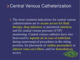
Central Venous Lines and Thoracic Drainage. Division of Cardiothoracic Surgery UWI Mona. Central Venous Lines: Why?. To monitor / ascertain hydration status To administer fluids / nutrition To administer medications To gain access to the right side of the heart and pulmonary arterial tree.

E N D
Central Venous Lines and Thoracic Drainage Division of Cardiothoracic Surgery UWI Mona
Central Venous Lines: Why? • To monitor / ascertain hydration status • To administer fluids / nutrition • To administer medications • To gain access to the right side of the heart and pulmonary arterial tree
Central venous lines can also be placed by gaining access from peripheral veins (basilic, cephalic, femoral) Peripherally-inserted central catheter “PICC” lines Access Sites, continued
Implantation Techniques • Sterile Technique • Seldinger Method Placement of needle inside vein. Guide wire through needle into vein, needle removed. Dilator over guide wire, dilator removed, wire left in vein. Catheter over guide wire into vein, guide wire removed, catheter left in situ. Safest, surest method of cannulation of vessels yet devised.
Open (Venesection) Catheter-thru-Needle Other Techniques:
Catheter-over-Needle Catheter-thru-cannula Other Techniques, con’td
Complications of CV lines • Incorrect puncture • Into tissues • With perforation of vessel (out the other side) • With arterial damage • Into pleural cavity pneumo, haemothorax • With nerve damage (brachial plexus)
Complications, con’td • Incorrect Catheter Position • In another vein (non-central, ascending) • Lumen openings outside venous lumen • Catheter positioned too far into right atrium incorrect pressure interpretation cardiac dysrhythmias cardiac perforation
Complications, con’td • Embolism • Catheter Embolism • Guidewire Embolism • Air Embolism Treatment may need to be immediate or adopt “wait and see” policy
Other Complications • Thrombosis • Local Infection • Local Haemorrhage • Disseminated Infection • (catheter-related septicaemia)
Hydration Status and Monitoring • In general: Central Venous Pressure 5 to 15 mmHg < 5 mmHg HYPOVOLEMIA > 15 mmHg HYPERVOLEMIA THIS IS ONLY TRUE FOR NORMAL, IDEALIZED INDIVIDUAL
Fluid Status / Monitoring • Ill patients are much more variable • An ill patient may be hypervolemic with a CVP of 10, or may be hypovolemic with a CVP of 16 • Use of the CV pressure monitoring line allows for close and precise evaluation of the patients’ intravascular volume status
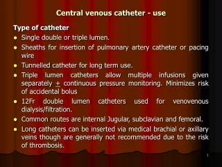
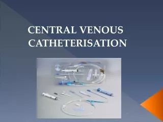
One or more (up to 3) lumina for simultaneous pressure monitoring and fluid / drug administration A Typical CV catheter:
Inserted into central vein via Seldinger technique, then advanced into pulmonary artery branch using pressure curves to guide placement Swan-Ganz Catheter
Swan-Ganz Catheter • When advanced into PA, blowing up of balloon gives information as to the LEFT ATRIAL PRESSURE, which relates directly to the filling pressure of the left ventricle, and from which detailed calculations of CARDIAC OUTPUT can be derived (thermodilution technique)
CV lines: Nutrition, Meds, Access to R. Heart • Nutrition: TPN (total parenteral nutrition) • Medications: Chemotherapy, vaso-sclerosant meds • Access: eg. Pacemaker wires Cardiac Biopsy
Chest Intubation(Tube Thoracostomy) : Why? • A procedure which becomes necessary when the pleural space contains air or fluid (blood, chyle, pus, bile, gastric contents, bowel contents, other fluids) in order that those abnormal contents can be drained and the underlying lung be allowed to re-expand to fill the pleural cavity
Tube Thoracostomy: When? • Following trauma (accidental, operative) • Following spontaneous pneumothorax • Following the development of a pus-filled chest after pneumonia or penetrating trauma (empyaema thoracis)
Chest Tube: How does it work? Fluid/Air drains into 1st bottle. Tube under H20 acts as a one-way valve fluid/air out, but not back in. Second bottle is a fluid trap. Third bottle allows for safe application of suction
Fluid in Pleural Space:Pus = Empyaema ThoracisBlood = HaemothoraxBlood + Air = Haemopneumothorax
Sites for ChestIntubation NB. Unusual sites are dictated by location of fluid collections
Incise • Make a linear incision along the rib, one interspace below the site of insertion of the tube
Dissect • Insert a curved clamp and tunnel superiorly to the interspace that is to be entered Avoid the neurovascular bundle at the inferior margin of the rib
Puncture • Gently but forcibly puncture through the intercostal muscles and parietal pleura. • A gush of air and/or blood will exit through the wound. • Avoid injuring the lung tissue when puncturing the pleura
Insertion • Insert a finger into the pleural space to identify and separate pleural adhesions With the clamp, grasp the tip of the chest tube and advance it into the thorax
Secure the tube • Direct the catheter posteriorly, superiorly • Secure the tube • Use an 0 silk suture into the skin through the incision. Come out through the opposite side. Then, wrap the suture around the tube several times and tie it securely in place.
Chest Tubes: Caveats • Excessively rapid drainage of fluid/air can result in too rapid re-expansion of ipsilateral lung leading to: RE-EXPANSION PULMONARY OEDEMA (not well understood) • NEVER clamp a chest tube, unless directed to do so by a chest surgeon!!
Conclusions • Central venous line insertion and chest intubation are two essential clinical skills • Both procedures are highly technical and may have complications, but modern techniques have greatly reduced adverse events