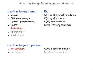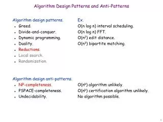
Intelligence and Patterns
Intelligence and Patterns. The Brain is a Pattern matching Machine. His book is free on line for download http://hubel.med.harvard.edu/index.html. PREFACE download 1 INTRODUCTION download 2 IMPULSES, SYNAPSES, AND CIRCUITS download


Intelligence and Patterns
E N D
Presentation Transcript
Intelligence and Patterns The Brain is a Pattern matching Machine
His book is free on line for download http://hubel.med.harvard.edu/index.html PREFACE download1 INTRODUCTIONdownload2 IMPULSES, SYNAPSES, AND CIRCUITSdownload 3 THE EYEdownload4 THE PRIMARY VISUAL CORTEX download5 THE ARCHITECTURE OF THE VISUAL CORTEXdownload6 MAGNIFICATION AND MODULESdownload7 THE CORPUS CALLOSUM AND STEREOPSISdownload8 COLOR VISION download9 DEPRIVATION AND DEVELOPMENTdownload10 PRESENT AND FUTUREdownloadFURTHER READINGdownload SOURCES OF ILLUSTRATIONS INDEX
Reverse-Engineering the BrainAt MIT, neuroscience and artificial intelligence are beginning to intersect. • www.technologyreview.com/read_article.aspx?id=17111 • While AI's progress has been slower than expected, neuro-science has gotten much more sophisticated in its understanding of how the brain works. Nowhere is this more obvious than in the 37 labs of MIT's BCS Complex. Groups here are charting the neural pathways of most of the higher cognitive functions (and their disorders), including learning, memory, the organization of complex sequential behaviors, the formation and storage of habits, mental imagery, number management and control, goal definition and planning, the processing of concepts and beliefs, and the ability to understand what others are thinking. • One breakthrough example: Biological vision solves problems in several different ways. One, according to Poggio's group, is to organize parallel processing around two simple operations and then alternate these operations in an ordered way through layers of neurons. Layer A might filter the basic inputs from the optic nerve; layer B would integrate the results from many cells in layer A; C would filter the inputs from B; D would integrate the results from C; and so on, perhaps a dozen times. As a signal rises through the layers, the outputs of the parallelized processors gradually combine, identity emerges, and noise falls away. • Some of their assumptions turned out to predict real features, such as the presence of cells (call them OR cells) that pick the strongest or most consistent signal out of a group of inputs and copy it to their own output fibers. (Imagine a group of three neurons, A, B, and C, all sending signals to OR neuron X. If those signals were at strength levels 1, 2, and 3 respectively, X would suppress A and B and copy C's signal to its output. If the strengths had been 3, 2, and 1, it would have copied A's signal and suppressed those of B and C.) • When human subjects and Serre and Poggio'simmediate-recognition program took the animal presence/absence test, the computer did as well as the humans -- and better than the best machine vision programs available. (Indeed, it got the right answer 82 percent of the time, while the humans averaged just 80 percent.)
Reverse Engineering the Brain • Processing without attention and consciousness • Rapid visual categorization • Visual input can be classified very rapidly. As famously demonstrated by Thorpe and colleagues (Thorpe et al., 1996; Kirchner and Thorpe, 2006) around 120 msec following image onset, some brain processes begin to respond differentially to images containing one of more animals from pictures than contain none. At this speed, it is no surprise that subjects often respond without having consciously seen the image; consciousness for the image may come later or not at all. • Dual-task and dual-presentation paradigms support the idea that such discriminations can occur in the near-absence of focal, spatial attention implying that purely feed-forward networks can support complex visual decision-making in the absence of both attention and consciousness. Indeed, this has now been formally shown in the context of a purely feed-forward computational model of the primate’s ventral visual system (Serre et al., 2007). www.scholarpedia.org/article/Attention_and_consciousness/processing_without_attention_and_consciousness www.technologyreview.com/printer_friendly_article.aspx?id=17111
One Example of Chunking Explaining Rapid Categorization. Thomas Serre, Aude Oliva, Tomaso Poggio.http://cbcl.mit.edu/seminars-workshops/workshops/serre-slides.pdf
Chunking Hierarchy The results by Logothetis et al. are in agreement with a general computational theory [Poggio, 2000] suggesting that a variety of visual object recognition tasks (involving the categorization of objects and faces at different levels) can be performed based on a linear combination of a few units tuned to specific task-related training examples. • The organization of visual cortex based on a core of knowledge that has been accumulated over the past 30 years. The figure is modified from[Oramand Perrett, 1994]mostly to include the likely involvement of prefrontal cortex during recognition tasks by setting task-specific circuits to read-out shape information from IT [Scalaidhe et al., 1999; Freedman et al., 2002, 2003; Hung et al., 2005]. Thomas Serre (2006), Learning a Dictionary of Shape-Components in Visual Cortex: Comparison with Neurons, Humans and Machines, Ph.D. dissertation, Department of Brain and Cognitive Sciences, Massachusetts Institute of Technology, April, http://cbcl.mit.edu/publications/ps/MIT-CSAIL-TR-2006-028.pdf
Cortical columnRetrieved from "http://en.wikipedia.org/wiki/Cortical_column" • A cortical column, also called hypercolumn or sometimes cortical module,[1] is a group of neurons in the braincortex which can be successively penetrated by a probe inserted perpendicularly to the cortical surface, and which have nearly identical receptive fields. Neurons within a minicolumn encode similar features, whereas a hypercolumn "denotes a unit containing a full set of values for any given set of receptive field parameters"[2]. A cortical module is defined as either synonymous with a hypercolumn (Mountcastle) or as a tissue block of multiple overlapping hypercolumns (Hubel&Wiesel). • Human cerebral cortex • The human cerebral cortex is composed of 6 somewhat distinct layers; each layer identified by the nerve cell type and the destination of these nerve cell's axons (within the brain). The human cortex is a roughly 2.4 mm thick sheet of neuronal cell bodies that forms the external surface of the telencephalon. The dolphin cortical column is composed of only 5 layers. The reptilian cortex has only three layers. • The columnar functional organization, as originally framed by Vernon Mountcastle, suggests that neurons that are horizontally more than 0.5 mm (500 µm) from each other do not have overlapping sensory receptive fields, and other experiments give similar results: 200–800 µm (Buxhoeveden 2002, Hubel 1977, Leise 1990, etc.). Various estimates suggest there are 50 to 100 cortical minicolumns in a hypercolumn, each comprising around 80 neurons. • An important distinction is that the columnar organization is functional by definition, and reflects the local connectivity of the cerebral cortex. Connections "up" and "down" within the thickness of the cortex are much denser than connections that spread from side to side. • Hubel and Wiesel studies • Hubel and Wiesel followed up on Mountcastle's discoveries in the somatic sensory cortex with their own studies in vision. A part of the discoveries that resulted in them winning the 1981 Nobel Prize[3] was that there were cortical columns in vision as well, and that the neighboring columns were also related in function in terms of the orientation of lines that evoked the maximal discharge. Hubel and Wiesel followed up on their own studies with work demonstrating the impact of environmental changes on cortical organization, and the sum total of these works resulted in their Nobel Prize. • Size of cortex • From the size of the cortex and the typical size of a column, it can be estimated that there are about two million function columns in humans [4]. There may be more if the columns can overlap, as suggested by Tsunodaet al[5]. • References • Kolb, Bryan; Whishaw, Ian Q. (2003). Fundamentals of human neuropsychology. New York: Worth. ISBN 0-7167-5300-6. • Horton JC, Adams DL (2005). "The cortical column: a structure without a function". Philos. Trans. R. Soc. Lond., B, Biol. Sci. 360 (1456): 837–62. doi:10.1098/rstb.2005.1623. PMID 15937015. • "The Nobel Prize in Medicine 1981". http://nobelprize.org/medicine/laureates/1981/. Retrieved on 2008-04-13. • Christopher Johansson and Anders Lansner (January 2007). "Towards cortex sized artificial neural systems". Neural Netw 20 (1): 48–61. doi:10.1016/j.neunet.2006.05.029. PMID 16860539. • Kazushige Tsunoda, Yukako Yamane, Makoto Nishizaki, and Manabu Tanifuji (August 2001). "Complex objects are represented in macaque inferotemporal cortex by the combination of feature columns". Nat. Neurosci. 4 (8): 832–8. doi:10.1038/90547. PMID 11477430.
Cortical minicolumnRetrieved from "http://en.wikipedia.org/wiki/Cortical_minicolumn" • A cortical minicolumn is a vertical column through the cortical layers of the brain, comprising perhaps 80–120 neurons, except in the primateprimary visual cortex (V1), where there are typically more than twice the number. There are about 2×10⁸minicolumns in humans.[1] From calculations, the diameter of a minicolumn is about 28–40 µm. • Many sources support the existence of minicolumns, especially Mountcastle,[2] with strong evidence reviewed by Buxhoeveden and Casanova[3] who conclude "... the minicolumn must be considered a strong model for cortical organization" and "[the minicolumn is] the most basic and consistent template by which the neocortex organizes its neurones, pathways, and intrinsic circuits". See also Calvin's Handbook on cortical columns. • Size – The minicolumn measures of the order of 40–50 µm in transverse diameter (Mountcastle 1997, Buxhoeveden 2000, 2001); 35–60 µm (Schlaug, 1995, Buxhoeveden 1996, 2000, 2001); 50 µm with 80 µm spacing (Buldyrev, 2000), or 30 µm with 50 µm (Buxhoeveden, 2000). Larger sizes may not be of human minicolumns, for example Macaque monkey V1 minicolumns are 31µm diameter, with 142 pyramidal cells (Peters, 1994) — 1270 columns per mm2. Similarly, the cat V1 has much bigger minicolumns, ~56 µm (Peters 1991, 1993) . • The size can also be calculated from area considerations: if cortex (both hemispheres) is 1.27×1011 µm2 then if there are 2×10⁸minicolumns in the cortex then each is 635 µm2, giving a diameter of 28 µm (if the cortex area were doubled to the commonly quoted value, this would rise to 40 µm). Johansson and Lansner[4] do a similar calculation and arrive at 36 µm (p51, last para). • Facts • Cells in 50µm minicolumn all have the same receptive field; adjacent minicolumns may have very different fields (Jones, 2000). • Downwards projecting axons in minicolumns are ≈10µm in diameter, periodicity and density similar to those within the cortex, but not necessarily coincident (DePhilipe, 1990). • Thalamic input (1 axon) reaches 100–300 minicolumns. • The number of fibres in the corpus callosum is 2–5×10⁸ (Cook 1984, Houzel 1999) — perhaps related to the number of minicolumns. • References • Towards cortex sized artificial neural systems, Christopher Johansson and Anders Lansner, Neural Networks, Vol. 20 #1, pp48–61, Elsevier, January 2007 • The columnar organization of the neocortex, Vernon B. Mountcastle, Brain, Vol. 20 #4, pp701–722, Oxford University Press, April 1997 • The minicolumn hypothesis in neuroscience, Daniel P. Buxhoeveden and Manuel F. Casanova, Brain, Vol. 125 #5, pp935–951, Oxford University Press, May 2002. • Towards cortex sized artificial neural systems, Christopher Johansson and Anders Lansner, Neural Networks, Vol. 20 #1, pp48–61, Elsevier, January 2007
Involuntary maybe, but certainly not random12Feb09 – www.physorg.com/news153670434.html • Our eyes are in constant motion. Even when we attempt to stare straight at a stationary target, our eyes jump and jiggle imperceptibly. Although these unconscious flicks, also known as microsaccades, had long been considered mere "motor noise," researchers at the Salk Institute for Biological Studies found that they are instead actively controlled by the same brain region that instructs our eyes to scan the lines in a newspaper or follow a moving object. • Their findings, published in the Feb. 13, 2009 issue of Science, provide new insights into the importance of these movements in generating normal vision. • "For several decades, scientists have debated the function, if any, of these fixational eye movements," says Richard Krauzlis, Ph.D., an associate professor in the Salk Institute's Systems Neurobiology Laboratory, who led the current study. "Our results show that the neural circuit for generating microsaccades is essentially the same as that for voluntary eye movements. This implies that they are caused by the minute fluctuations in how the brain represents where you want to look." • "There was a lot of past effort to figure out what fixational eye movements contribute to our vision," adds lead author Ziad Hafed, Ph.D., Sloan-Swartz Fellow in the Systems Neurobiology Laboratory, "but nobody had looked at the neural mechanism that generates these movements. Without such knowledge, one could only go so far in evaluating microsaccades' significance and why they actually exist." • Wondering whether the command center responsible for generating fixational eye movements resides within the same brain structure that is in charge of initiating and directing large voluntary eye movements, Hafed decided to measure neural activity in the superior colliculus before and during microsaccades. • He not only discovered that the superior colliculus is an integral part of the neural mechanism that controls microsaccades, but he also found that individual neurons in the superior colliculus are highly specific about which particular microsaccade directions and amplitudes they command—whether they be, say, rightward or downward or even oblique movements. "Data from the population of neurons we analyzed shows that the superior colliculus contains a remarkably precise representation of amplitude and direction down to the tiniest of eye movements," says Krauzlis. • The Salk researchers, in collaboration with Laurent Goffart, Ph.D., a professor at the Institut de Neurosciences Cognitives de la Méditerranée in Marseille, France, also temporarily inactivated a subset of superior colliculus neurons and analyzed the resulting changes in microsaccades. They discovered that a fully functional superior colliculus is required to generate normal microsaccades. • "Because images on the retina fade from view if they are perfectly stabilized, the active generation of fixational eye movements by the central nervous system allows these movements to constantly shift the scene ever so slightly, thus refreshing the images on our retina and preventing us from going 'blind,'" explains Hafed. "When images begin to fade, the uncertainty about where to look increases the fluctuations in superior colliculus activity, triggering a microsaccade," adds Krauzlis. • Microsaccades may, however, do more than prevent the world around us from fading when we stare at it for too long. Even when our gaze is fixed, our attention can shift to an object at the periphery that attracts our interest. In an earlier study, Hafed discovered that although we may avert our eyes from an attractive man or woman, microsaccades will reveal such objects of attraction because their direction is biased toward objects to which we are unconsciously attracted. • By showing in the current study that the superior colliculus is involved in generating microsaccades, Hafed and his colleagues could now explain why this happens. "The superior colliculus is a major determinant of what is behaviorally relevant in our visual environment, so paying attention to one location or the other alters superior colliculus activity and therefore alters these eye movements as well," says Hafed.
The Blue Brain project is the first comprehensive attempt to reverse-engineer the mammalian brain, in order to understand brain function and dysfunction through detailed simulations. • In July 2005, EPFL and IBM announced an exciting new • research initiative - a project to create a biologically accurate, functional model of the brain using IBM's Blue Gene supercomputer. Analogous in scope to the Genome Project, the Blue Brain will provide a huge leap in our understanding of brain function and dysfunction and help us explore solutions to intractable problems in mental health and neurological disease. • At the end of 2006, the Blue Brain project had created a model of the basic functional unit of the brain, the neocortical column. At the push of a button, the model could reconstruct biologically accurate neurons based on detailed experimental data, and automatically connect them in a biological manner, a task that involves positioning around 30 million synapses in precise 3D locations. • In November, 2007, the Blue Brain project reached an important milestone and the conclusion of its first Phase, with the announcement of an entirely new data-driven process for creating, validating, and researching the neocortical column.
About the Blue Brain Projecthttp://bluebrain.epfl.ch/page18699.html • The cerebral cortex, the convoluted "grey • matter" that makes up 80% of the human • brain, is responsible for our ability to • remember, think, reflect, empathize, communicate, adapt to • new situations and plan for the future. The cortex first • appeared in mammals, and it has a fundamentally simple • repetitive structure that is the same across all mammalian • species. • The brain is populated with billions of neurons, each connected to thousands of its neighbors by dendrites and axons, a kind of biological "wiring". The brain processes information by sending electrical signals from neuron to neuron along these wires. In the cortex, neurons are organized into basic functional units, cylindrical volumes 0.5 mm wide by 2 mm high, each containing about 10,000 neurons that are connected in an intricate but consistent way. These units operate much like microcircuits in a computer. This microcircuit, known as the neocortical column (NCC), is repeated millions of times across the cortex. The difference between the brain of a mouse and the brain of a human is basically just volume - humans have many more neocortical columns and thus neurons than mice. • This structure lends itself to a systematic modeling approach. And indeed, the first step of the Blue Brain project is to re-create this fundamental microcircuit, down to the level of biologically accurate individual neurons. The microcircuit can then be used in simulations. • For an in-depth view of the project, read Henry Markram's Perspectives article in the February 2006 issue of Nature Reviews Neuroscience.
Building the microcircuithttp://bluebrain.epfl.ch/page19092.html • Modeling Neurons – Neurons are not all alike - they come in a • variety of complex shapes. The precise shape and structure of a • neuron influences its electrical properties and connectivity with • other neurons. A neuron's electrical properties are determined to a large extent by a • variety of ion channels distributed in varying densities throughout the cell's • membrane. Scientists have been collecting data on neuron morphology and electrical • behavior of the juvenile rat in the laboratory for many years, and this data is used as • the basis for a model that is run on the Blue Gene to recreate each of the 10,000 • neurons in the NCC. • Modeling connections – To model the neocortical column, it is essential to • understand the composition, density and distribution of the numerous cortical cell types. Each class of cells is present in specific layers of the column. The precise density of each cell type and the volume of the space it occupies provides essential information for cell positioning and constructing the foundation of the cortical circuit. Each neuron is connected to thousands of its neighbors at points where their dendrites or axons touch, known as synapses. In a column with 10,000 neurons, this translates into trillions of possible connections. The Blue Gene is used in this extremely computationally intensive calculation to fix the synapse locations, "jiggling" individual neurons in 3D space to find the optimal connection scenario. • Modeling the column – The result of all these calculations is a re-creation, at the cellular level, of the neocortical column, the basic microcircuit of the brain. In this case, it's the cortical column of a juvenile rat. This is the only biologically accurate replica to date of the NCC - the neurons are biologically realistic and their connectivity is optimized. This would be impossible without the huge computational capacity of the Blue Gene. A model of the NCC was completed at the end of 2006. • In November, 2007, The Blue Brain Project officially announced the conclusion of Phase I of the project, with three specific achievements: • A new modeling framework for automatic, on-demand construction of neural circuits built from biological data • A new simulation and calibration process that automatically and systematically analyzes the biological accuracy and consistency of each revision of the model • The first cellular-level neocortical column model built entirely from biological data that can now serve as a key tool for simulation-based research