Download
1 / 32
480 likes | 1.04k Vues
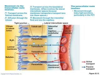
Movement via the transcellular route involves:. The paracellular route involves:. Transport across the basolateral membrane. (Often involves the lateral intercellular spaces because membrane transporters transport ions into these spaces.). 3. • Movement through

E N D
Movement via the transcellular route involves: The paracellular route involves: Transport across the basolateral membrane. (Often involves the lateral intercellular spaces because membrane transporters transport ions into these spaces.) 3 • Movement through leaky tight junctions, particularly in the PCT. 1 Transport across the luminal membrane. 4 Diffusion through the cytosol. Movement through the interstitial fluid and into the capillary. 2 Lateral intercellular space Tight junction Filtrate in tubule lumen Interstitial fluid Tubule cell Peri- tubular capillary Capillary endothelial cell Paracellular H2O 2 3 4 1 Transcellular Luminal membrane 1 3 4 Transcellular 2 Solutes 3 4 Active transport Paracellular Basolateral membranes Passive transport Figure 25.13
Reabsorption of Nutrients, Water, and Ions • Na+ reabs provides means for reabsorbing most other substances • Organics are reabsby secondary active transport • Transport maximum (Tm) reflects number of carriers in renal tubules available • When carriers are saturated, excess of that substance is excreted
Reabsorption of Nutrients, Water, and Ions • Water is reabsby osmosis (obligatory water reabsorption), aided by water pores called aquaporins-ADH • Cations and fat-soluble substances follow by diffusion
1 At the basolateral membrane, Na+ is pumped into the interstitial space by the Na+-K+ ATPase. Active Na+ transport creates concentration gradients that drive: Nucleus Filtrate in tubule lumen Interstitial fluid Peri- tubular capillary 2 Tubule cell “Downhill” Na+ entry at the luminal membrane. 3 Reabsorption of organic nutrients and certain ions by cotransport at the luminal membrane. Na+ 2 3Na+ 3Na+ 1 Glucose Amino acids Some ions Vitamins 2K+ 2K+ 4 Reabsorption of water by osmosis. Water reabsorption increases the concentration of the solutes that are left behind. These solutes can then be reabsorbed as they move down their concentration gradients: 3 K+ 4 H2O 5 Lipid-soluble substances 5 Lipid-soluble substances diffuse by the transcellular route. 6 Cl–, Ca2+, K+ and other ions, urea Cl– Paracellular route Tight junction 6 Cl– (and other anions), K+, and urea diffuse by the paracellular route. Transport protein Primary active transport Secondary active transport Ion channel or aquaporin Passive transport (diffusion) Figure 25.14
Reabsorptive Capabilities of Renal Tubules and Collecting Ducts • Mechanism of aldosterone • Targets collecting ducts and DCT • Promotes synthesis of Na+ and K+channels
Regulation of Urine Concentration and Volume • Osmolality • Number of solute particles in 1 kg of H2O • Reflects ability to cause osmosis
Regulation of Urine Concentration and Volume • Osmolality of body fluids • Expressed in milliosmols (mOsm) • The kidneys maintain osmolality of plasma at ~300 mOsm,
Countercurrent Mechanism • Occurs when fluid flows in opposite directions in two adjacent segments of the same tube • Filtrate flow in the loop of Henle (countercurrent multiplier) • Blood flow in the vasa recta (countercurrent exchanger)
Countercurrent Mechanism • Role of countercurrent mechanisms • Establish and maintain an osmotic gradient (300 mOsm to 1200 mOsm) from renal cortex through the medulla • Allow the kidneys to vary urine concentration
Cortex Medulla Figure 25.15
Formation of Concentrated Urine • Depends on medullary osmotic gradient and ADH • ADH triggers reabof H2O in collecting ducts • Facultative water reabsorption occurs in presence of ADH so that 99% of H2O in filtrate is reabs
Diuretics • Chemicals that enhance the urinary output • Osmotic diuretics: substances not reabsorbed, (e.g., high glucose in a diabetic patient) • ADH inhibitors such as alcohol • Substances that inhibit Na+reabsand obligatory H2O reabsorptionie.caffeine and many drugs
Milliosmols Na+ (65%) Glucose Amino acids H2O (65%) and many ions (e.g. Cl– and K+) Cortex (d) (a) 300 (e) Outer medulla (b) (c) 600 Some drugs HCO3– H+, NH4+ Inner medulla Blood pH regulation (a) Proximal convoluted tubule: • 65% of filtrate volume reabsorbed • Na+, glucose, amino acids, and other nutrients actively transported; H2O and many ions follow passively • H+ and NH4+ secretion and HCO3– reabsorption to maintain blood pH (see Chapter 26) • Some drugs are secreted 1200 Active transport (primary or secondary) Passive transport Figure 25.18a
Milliosmols Cortex (d) (a) 300 (e) Outer medulla H2O (b) (c) 600 (b) Descending limb of loop of Henle • Freely permeable to H2O • Not permeable to NaCl • Filtrate becomes increasingly concentrated as H2O leaves by osmosis Inner medulla 1200 Active transport (primary or secondary) Passive transport Figure 25.18b
Milliosmols Cortex Na+ (d) Cl– (a) K+ 300 (e) Outer medulla (b) Urea Na+ (c) Cl– 600 Inner medulla (c) Ascending limb of loop of Henle • Impermeable to H2O • Permeable to NaCl • Filtrate becomes increasingly dilute as salt is reabsorbed 1200 Active transport (primary or secondary) Passive transport Figure 25.18c
Na+; aldosterone-regulated Ca2+; PTH-regulated Cl–; follows Na+ Milliosmols Cortex (d) (a) 300 (e) Outer medulla (b) (c) 600 Inner medulla (d) Distal convoluted tubule • Na+ reabsorption regulated by aldosterone • Ca2+ reabsortion regulated by parathyroid hormone (PTH) • Cl– cotransported with Na+ 1200 Active transport (primary or secondary) Passive transport Figure 25.18d
Milliosmols Cortex H2O regulated by ADH (d) (a) 300 Regulated by aldosterone: Urea; increased by ADH (e) Na+ Outer medulla (b) K+ Blood pH regulation (c) 600 H+ HCO3– Inner medulla NH4+ 1200 (e) Collecting duct • H2O reabsorption through aquaporins regulated by ADH • Na+ reabsorption and K+ secretion regulated by aldosterone • H+ and HCO3– reabsorption or secretion to maintain blood pH (see Chapter 26) • Urea reabsorption increased by ADH Active transport (primary or secondary) Passive transport Figure 25.18e
Renal Clearance • Volume of plasma cleared of a particular substance in a given time • Renal clearance tests are used to • Determine GFR • Detect glomerular damage
Renal Clearance RC = UV/P RC = renal clearance rate (ml/min) U = concentration (mg/ml) of substance in urine V = flow rate of urine formation (ml/min) P = conc of same substance in plasma
Renal Clearance • For any substance freely filtered and neither reabsorbed nor secreted by the kidneys (e.g., insulin), RC = GFR = 125 ml/min • If RC < 125 ml/min, substance is reabsorbed • If RC = 0, substance is completely reabsorbed • If RC > 125 ml/min, substance is secreted (most drug metabolites) • Duck vs Beaver
Lumen Adventitia Circular layer Longitudinal layer Transitional epithelium Lamina propria Figure 25.20
Renal Calculi • Kidney stones form in renal pelvis • Crystallized calcium, magnesium, or uric acid salts • Larger stones block ureter, cause pressure and pain in kidneys • May be due to chronic bacterial infection, urine retention, Ca2+ in blood, pH of urine
Urinary Bladder • Trigone • Triangular area outlined by openings for ureters and urethra • Infections tend to persist in this region
Urinary Bladder • Layers of the bladder wall • Transitional epithelial mucosa • Thick detrusor muscle (three layers of smooth muscle) • Fibrous adventitia (peritoneum on superior surface only)
Urinary Bladder • Collapses when empty; rugae appear • Expands and rises superiorly during filling without significant rise in internal pressure
Peritoneum Ureter Rugae Detrusor muscle Ureteric orifices Bladder neck Internal urethral sphincter Trigone External urethral sphincter Urogenital diaphragm Urethra External urethral orifice (b) Female. Figure 25.21b
Micturition • Urination or voiding • Three simultaneous events • Contraction of detrusor muscle by ANS • Opening of internal urethral sphincter by ANS • Opening of external urethral sphincter by somatic nervous system
Brain Higher brain centers Urinary bladder filling stretches bladder wall Allow or inhibit micturition as appropriate Pontine micturition center Pontine storage center Afferent impulses from stretch receptors Promotes micturition by acting on all three spinal efferents Inhibits micturition by acting on all three spinal efferents Simple spinal reflex Spinal cord Spinal cord Parasympathetic activity Sympathetic activity Somatic motor nerve activity Parasympathetic activity Sympathetic activity Somatic motor nerve activity Detrusor muscle contracts; internal urethral sphincter opens External urethral sphincter opens Inhibits Micturition Figure 25.22