Download
1 / 29
290 likes | 455 Vues
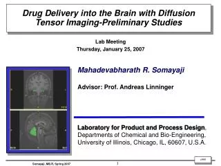
Drug Delivery into the Brain with Diffusion Tensor Imaging-Preliminary Studies. Lab Meeting Thursday, January 25, 2007. Mahadevabharath R. Somayaji Advisor: Prof. Andreas Linninger Laboratory for Product and Process Design ,

E N D
Drug Delivery into the Brain with Diffusion Tensor Imaging-Preliminary Studies Lab Meeting Thursday, January 25, 2007 Mahadevabharath R. Somayaji Advisor: Prof. Andreas Linninger Laboratory for Product and Process Design, Departments of Chemical and Bio-Engineering, University of Illinois, Chicago, IL, 60607, U.S.A.
Motivation • Predicting the fate of drugs for the treatment of neurodegenerative diseases in the human brain is still elusive. • Better understanding the transport mechanism of therapeutic drugs in human brain tissue using diffusion tensor imaging data. • 2. Calculation of achievable treatment volumes in a patient-specific three dimensional human brain using first principles. • 3. Investigating the role and choice of infusion catheter in convection enhanced drug delivery
Presentation Overview • Demonstration of the Diffusion Phenomenon • Two-dimensional Results • 2.1. Filling DTI data for 2D Mesh Files • 2.2 Scaling Procedure • 2.3 Steady State Results: Drug Distribution using Multiple Hole Catheter • 2.4 Dynamic Results: Drug Distribution using Multiple Hole Catheter in four weeks • 2.4.1. Comparison of Achievable Treatment Volumes • Three-dimensional Results • 3.1 Dynamic Results: Drug distribution in the brain four weeks after shown in plane – • projections • Remaining Work • Acknowledgements
Demonstration of Diffusion Phenomenon Anisotropic, Homogeneous Isotropic, Homogeneous Anisotropic, Homogeneous Anisotropic, Homogeneous_v2 Anisotropic, Inhomogeneous
ImageJ *.raw x y y x Filling the DTI Data for 2D Mesh Files Gambit *.msh MRI *.raw Eigenvector Check! 1 3 2 3 1 2 2 3 1
Filling the DTI Data into the Mesh File # volumes = 648 # cell centroids = 648 Vol 612 Vol 615 Vol 636 Vol 576
Visualization of Main Diffusivities from Diffusion Tensor Imaging (DTI) Fiber Tract orientation Fractional Anisotropy Map Eigenvector Map
Visualization of Main Diffusivities from Diffusion Tensor Imaging (DTI) Fractional Anisotropy Map
Transport Phenomena in the brain: Theory and Mathematical Background Drug Transport Drug Transport porous brain parenchyma Effective Diffusivity in Porous Tissue Prediction of Bulk Diffusivity of Drug Effective Diffusion Tensor Bulk Flow Continuity Equation Equation of Motion Additional pressure drop in porous tissue Hydraulic Conductivity Tensor
Raw DTI data 1 Tensor Analysis 2 Determine: 1. Effective Diffusion tensor of drug 2. Permeability tensor of tissue 3. Inertial Parameter tensor of tissue Profile filter 3 Generate Profile files 4 FLUENT Software Scaling Procedure to obtain Drug Diffusion Tensor and Tissue Permeability Tensor Sharing of Eigenvectors References: 1. Sarntiroranont, M; Chen, X; Zhao, J, and Mareci, T.M, “Computational Model of Interstitial Transport in the Spinal Cord using Diffusion Tensor Imaging”, Annals of Biomedical Engineering, 34(8), pp. 1304-1321, 2006. 2. Tuch, D.S;.Weeden, V.J; Dale, A.M; George, J.S, and Belliveau, W, “Conductivity Tensor Mapping of the Human Brain using Diffusion Tensor MRI, Proc. Nat. Acad. Sci, 98, pp. 11697-11701, 2001.
2 Eigenvalue Decomposition to form FULL Tensor (i) SCALING: Diffusion tensor of drug (ii) SCALING: Permeability of the tissue Eigenvalue Normalization Permeability: Baseline Scaling Parameter (iii) SCALING: Inertial Parameter of the tissue Bulk Diffusivity: Baseline Scaling Parameter
Boundary Conditions Coronal section Axial section Ventricles Injection site Brain Tissue Pia-arachnoid membrane
Anisotropic-Inhomogeneous Diffusion in an Axial Human Brain Section: Steady State Results Diffusion Only Injection Site #2 Injection Site #1 Concentration field Low-flow Infusion Pressure field Flow field Concentration field
Multiple Hole Catheter Catheter tip Multiple Hole Catheter Brain Tissue Catheter Shaft # of holes on each side: 10 Outer diameter of each hole: 0.05 mm Mesh Info: # of volumes: 80336
Multiple Hole Catheters: Results- four weeks after # of holes on each side: 10 Total # of holes: 20 Outer diameter of each hole: 0.05 mm Constant infusion rate = 6 μl/min Flow field near the outlet Pressure Drug release from multiple holes
Comparison of Achievable Treatment Volumes between Single and Multiple hole Catheters four weeks after Infusion mode: Constant infusion (Low-flow microinfusion) Flow Rate: 6 μl/min Drug: Trophic Factor
Three dimensional computational mesh of the human brain Infusion Parameters Infusion Type: Constant Flow Rate: 6 μl/min Drug: Trophic Factor Catheter Diameter: 1.0 mm Catheter Type: Single hole Infusion Site: Near Putamen Mesh Info: # of tetrahedral volumes: 280438
Illustration of Two-Dimensional Projections from a 3D Volume Axial Sagittal Coronal 3D Brain
Distribution of drug in three dimensions shown in plane projections: Results four weeks after Coronal cut Axial cut Sagittal cut
Distribution of drug in three dimensions shown in plane projections: Results four weeks after
Distribution of drug in three dimensions shown in plane projections: Results four weeks after
Remaining Work • Pulsed Injection • Intracerebroventricular Infusion • Validate and finalize multiple hole catheter • 3D: Finalize dynamic simulation • Comparison of computational results with experimental data
Acknowledgements • Prof. Andreas Linninger, Director, LPPD, UIC • Dr. Libin Zhang, LPPD, UIC • Dr. Richard Penn, M.D, Univ. of Chicago • Dr. Guo, Univ. of Chicago • Dr. Michalis Xenos, LPPD, UIC • Monil Shah, LPPD, UIC