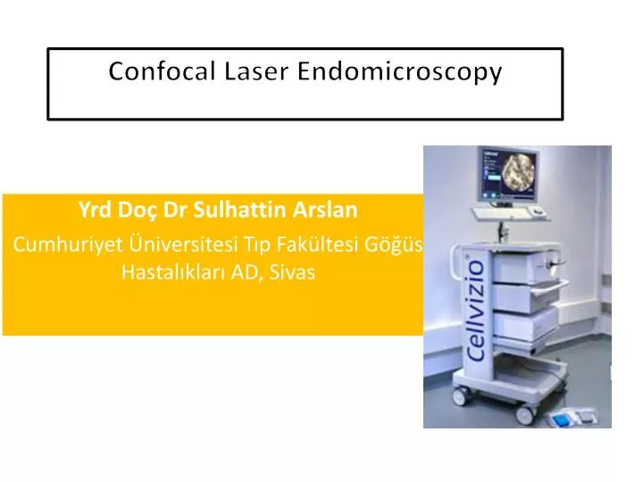

Confocal Laser Endomicroscopy
E N D
Presentation Transcript
ConfocalLaserEndomicroscopy YrdDoçDrSulhattin Arslan Cumhuriyet Üniversitesi Tıp Fakültesi Göğüs Hastalıkları AD, Sivas
HERHANGİ BİR KURUM YA DA ŞAHIS İLE BİR ÇIKAR İLİŞKİM YOKTUR
ConfocalLaserEndomicroscopy • Confocal endomicroscopes aim at providing microscopic imaging of a living tissue to the clinician • ‘‘Optical biopsies’’
ConfocalLaserEndomicroscopy • Such systems have recently been applied to the explorations of several organs, including the upper and the lower GIS, pancreato-biliary tract • More recently to the proximal and distal airways in vivo
ConfocalLaserEndomicroscopy 1- Optisan/Pentax Microendoscopy,Japan 2- CellVizio-LungR /Mauna Kea Tech, French Commercially;
Optisan/PentaxEndomicroscopy Optiscan endomicroscopy of the gastrointestinal tract provide imagesvery close to conventional histology
Optisan/PentaxEndomikroskopi Two potential drawbacks explainwhy this system is not yet available for the respiratory tractimaging; Largediameter of distal Scanning rate is tooslow (1 frame/second) Respiratorysystem is more mobile
CellVizio-LungR • A probe-based confocal laser endomicroscopy • Designed for imaging of the internal micro-structures of anatomical tissues
CellVizio-LungR The device has a laser scanning technology adapted to image by means of an optical fiber bundle
CellVizio-LungR Uses the principle of proximal scanning Carriesthelighttothescannedareaby fiber bundleandminiprobe Thelight delivery, scanning, spectral filtering, and imaging systemsare located at the proximal part of the device Thedistalpartincludes a connectorwhichtransferstheimagetotheminiprobeandthelaserscanningunit
ConfocalLaserEndomicroscopy • Confocalmicroscopyhelpstoimage in vivocellsandtissuesbyobtaininglateralandaxialresolution • Theilluminationanddetectionsystems being conjugated on the same focal plane, aretermedconfocal
ConfocalLaserEndomicroscopy • 488 nm wavelength is used for imaging • Microspectrometer experiments have clearly demonstrated thatthe main fluorescence signal emitted after 488 nm excitationfrom both bronchial and alveolar human systems originates fromthe elastin component of the tissue • Elastin fibers
ConfocalLaserEndomicroscopy • 1.4 nm diameter miniprobe • 488-nm-wavelength laser light • 9-12 frames per second • Depth of focus of 0 to 50 micron • Field of view of 600 3 600 micron • Lateral resolution of 3.5 μm • Up to 30,000 microfibers Histological quality image
MiniprobE • The miniprobe is applied onto thebronchial wall surface or advanced into a distal bronchiole down tothe acinusandobtainsimagesfromtheseanatomicalareas. Thebasementmembrane of the proximal airways Peripheralinterstitial Alveolarwall Macrophage Bronchialepitheliallayer Peripherallungnodules
ConfocalLaserEndomicroscopy Alveolar septal wall Microvessels Macrophage Basement membrane
ConfocalLaserEndomicroscopy • After the procedure Hemorrhage (because of miniprobe) there is no need to intervention
ConfocalLaserEndomicroscopy • Contrast agent Intravenous: Fluorescein %1,4 emesis, hypotension and epigastric pain Topical: Acriflavine, Metilen blue
Imaging of the Proximal Bronchi • Underlocalanesthesia • Miniprobe is introducedinto theworking channel of the bronchoscope • The depth of focus being 50 mm below the contactsurface, subepithelialconnectivetissue lamina densa, laminareticularis 480 nmfluoreskeine bronchialbasementmembrane Benign, malignant/premalignantbronchialalterations
FromtheDistalBronchioles Down to the Lung Acini Elastin represents up to 50% of the peripheral lung connective tissue fibers Bronchial wall, alveolus, extra-alveolar microvessels
Confocallaserendomicroscopyfordiagnosinglungcancer in vivo 32 patients with suspected malignancies underwent bronchoscopy with endomicroscopy using acriflavine 75,522 confocalimagesfrom 56 differentlocations Neoplasticchanges: Sensitivity, 96% Specificity, 87% Accuracy, 91% Fuchs et al. EurRespir J. 2012 Sep 20. [Epub ahead of print]
ConfocalLaserEndomicroscopy • Peripheral pulmonary nodule • Interstitial pathology • Bronchi and bronchial structure • Chronic rejection after lung transplantation
limitations • Can not obtaintissuesample • Onlyelastin fiber image