Download
1 / 38
380 likes | 605 Vues
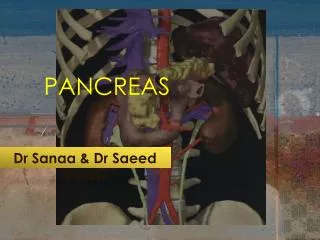
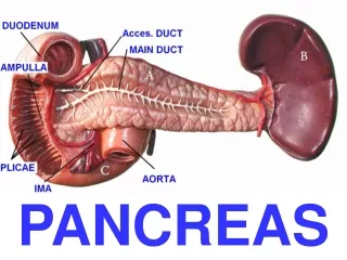
Pancreas and L iver. Pancreas. The pancreas is a mixed exocrine-endocrine gland that produces both digestive enzymes and hormones A thin capsule of connective tissue covers the pancreas and sends septa into it, separating the pancreatic lobules.

E N D
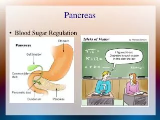
Pancreas • The pancreas is a mixed exocrine-endocrine gland that produces both digestive enzymes and hormones • A thin capsule of connective tissue covers the pancreas and sends septa into it, separating the pancreatic lobules. • The secretoryacini are surrounded by a basal lamina that is supported by a delicate sheath of reticular fibers and a rich capillary network.
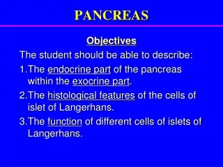
The digestive enzymes are produced by cells of the larger exocrine portion. • and the hormones are synthesized in clusters of endocrine epithelial cells known as pancreatic islets (islets of Langerhans). • The exocrine portion of the pancreas is a compound acinar gland, similar in structure to the parotid gland. • The two glands can be distinguished histologically by the absence of striated ducts and the presence of the islets in the pancreas.
Another characteristic detail is that in the pancreas the initial portions of intercalated ducts penetrate the lumens of the acini. • Small pale-staining centroacinar cells constitute the intraacinar portion of the intercalated duct and are found only in pancreatic acini. • Intercalated ducts merge to form larger interlobular ducts lined by columnar epithelium. • No ducts in the pancreas are striated.
Each exocrine acinus of the pancreas is composed of several serous cells surrounding a very small lumen • The acinar cells are highly polarized, with a spherical nucleus, and are typical protein-secreting cells • The number of zymogen granules present in each cell varies and is maximal in animals that have fasted.
The exocrine pancreas secretes 1.5 to 2 L of fluid per day. • Pancreatic juice is rich in bicarbonate ions (HCO3–) and digestive enzymes, including several proteases (trypsinogens, chymotrypsinogen, proelastases, protease E, kallikreinogen, procarboxipeptidases), -amylase, lipases, and nucleases (DNAase and RNAase).
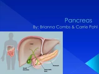
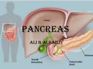
Liver • Except for the skin the liver is the body's biggest organ, weighing about 1.5 kg or about 2% of an adult's body weight. • With a large right lobe and smaller left lobe, it is the largest gland and is situated in the abdominal cavity beneath the diaphragm • The liver is an interface between the digestive system and the blood: the organ in which nutrients absorbed in the digestive tract are processed for use by other parts of the body.
Afferent vessel of liver • Most blood in the liver (70–80%) comes from the portal vein arising from the stomach, intestines, and spleen; the rest (20–30%) is supplied by the hepatic artery.
Portal vein • All the materials absorbed via the intestines reach the liver through the portal vein, except the complex lipids (chylomicrons), which are transported mainly by lymph vessels. • (portal vein – interlobar veins- interlobular veins-into hepatic sinusoids-central vein- sublobar vein-hepatic vein-inferior vena cava)
Hepatic artery • Brings arterial blood to liver. • Enters through portahepatis • Divide into interlobular arteries • drain into portal vein
Efferent vessel of liver • Hepatic vein which drain into inferior vena cava.
The position of the liver in the circulatory system is optimal for gathering, transforming, and accumulating metabolites from blood and for neutralizing and eliminating toxic substances in blood. • The elimination occurs in the bile, an exocrine secretion of the liver that is important for lipid digestion in the gut. • The liver also produces plasma proteins such as albumin, fibrinogen, and various carrier proteins.
Hepatocytes • Most of the hepatocytes have a single spherical nucleus • 25% of them are binucleated • Each nucleus has one or two nucleoli. • Contain cell organelles. • Mitochondria scattered through out. • Golgi apparatus are present in peripheral part. • Microsomes and peroxisomes are abundant. • Cytoplasm of hepatocytes stains acidophhillic (abundant mitochondria) and SER • Some areas of basophillic due to RER.
Contain large number of glycogen granules and fat droplets. In H & E these preparations, substances are washed out, leaving empty spaces in the cytoplasm. • Surface of hepatocytes is seperated from the wall of the sinusoid by a narrow space that contain type 3 collagen and blood plasma. This space is called perisinusoidal space (space of Disse)… space of exchange of substances between blood plasma and liver cells
Stroma • The liver is covered by a thin fibrous capsule of connective tissue (Glisson’s capsule)that becomes thicker at the hilum, where the portal vein and the hepatic artery enter the organ and where the right and left hepatic ducts and lymphatics exit. • These vessels and ducts are surrounded by connective tissue all the way to their termination (or origin) in the portal spaces between the liver lobules. • At this point, a delicate reticular fiber network surrounds and supports the liver cells and the sinusoidal endothelial cells of the liver lobules
Liver parenchyma • It consists of masses of epithelial cells called hepatocytes • Between the plates are present blood sinusoids called hepatic sinusoids • 3 liver structure have been proposed • 1. classic hepatic lobule • 2. portal lobule • 3. Hepatic acinus
Classic hepatic lobule • Shaped roughly like a polygonal prism, measuring about 0.7 mm in width and 2 mm in length • The central structure of the lobule traversing its long axis is a relatively large venule called central vein, which is a tributary of hepatic vein. • Plates are one cell thick and are seperated from each other by hepatic sinusoids. • The cross sections of hepatic lobules appear hexagonal in shape • Radiating plates of hepatocytes are seen as cords called hepatic cords.
At the angles of the hexagonal hepatic lobule are present roughly triangular areas called portal areas. Contain 3 tubular structures • Portal vein • Hepatic artery • Bile duct • Collectively called as portal triad.
Afferent blood vessels (portal vein and hepatic artery) at the corners • Efferent blood vessel (central vein) in centre so • Blood flows from periphery through the blood sinusoids to central vein. • On the other hand bile passes from liver cells to the bile duct lying at the periphery of the lobule • The bile flows in narrow channels called bile canaculi(spaces between adjacent liver cells)
Portal lobule • This is based on exocrine function of the liver i.e. production of bile • A portal lobule has at its centre a portal area • Triangular in cross section, contains parts of three adjoining hepatic lobules • Central vein at its three corners
Hepatic acinus(of Rappaport) • Area of liver parenchyma which surrounds a terminal branch of a portal vein, hepatic arteriole and bile ductule. • Diamond in cross section • Is situated in adjacent areas of two different classic hepatic lobules. • One central vein is located at each of the two opposite corners of the diamond shaped area • Terminal branches of portal vein and hepatic artery course transversely in the centre of the acinus.
Hepatic Lobules • Liver cells or hepatocytes (Gr. hepar, liver, + kytos, cell) are epithelial cells grouped in interconnected plates. • Hepatocytes are arranged into thousands of small (0.7 x 2 mm), polyhedral hepatic lobules which are the classic structural and functional units of the liver. • Each lobule has three to six portal areas at its periphery and a venule called a central vein in its center.
This exchange is the key to liver function, not only because of the large number of macromolecules (eg, lipoproteins, albumin, and fibrinogen) secreted into the blood by hepatocytes but also because the liver takes up and catabolizes many of these large molecules.
Liver sinusoids are surrounded and supported by delicate sheathes of reticular fibers • Two noteworthy cells are associated with these sinusoids in addition to the endothelial cells: • Ito cells • Kupffer cells
Kupffer cells • Abundant specialized sinusoidal macrophages, also known as Kupffer cells, are found between sinusoidal endothelial cells and on the luminal surface within the sinusoids, mainly near the portal areas. • Their main functions are to break down aged erythrocytes and free heme for re-use, remove bacteria or debris that may enter the portal blood from the gut, and act as antigen-presenting cells in adaptive immunity.
Ito cells • In the perisinusoidal space (not the lumen) are stellatefat storing cells (or Ito cells) with small lipid droplets containing vitamin A. • These cells, which make up about 8% of the cells in a liver but are difficult to see in routine preparations, store much of the body's vitamin A, produce ECM components, and have a regulatory role in local immunity. • In cirrhosis of liver these cells secrete type 1 and 2 collagen, deposited in the space of Disse, resulting in fibrosis of liver.