Download
1 / 27
270 likes | 474 Vues
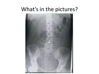
What’s in the pictures?. What’s in the pictures?. What’s in the pictures?. Medical Uses of X-rays and Ultrasound. How can we use X rays and Ultrasound to see inside people?. Starter : Why do we not use x rays to look at growing babies? . Glossary .

E N D
Medical Uses of X-rays and Ultrasound How can we use X rays and Ultrasound to see inside people? Starter: Why do we not use x rays to look at growing babies?
Glossary • Radiograph – the use of x-rays to look at non-uniform material e.g. human body. • Electromagnetic Spectrum – the range of frequencies of electromagnetic radiation.
Xrays • http://www.bbc.co.uk/learningzone/clips/medical-uses-of-x-rays-the-electromagnetic-spectrum/1455.html
How do they work? • X rays are directed at the patient who has a photographic film or flat-panel detector either side of them. • X-rays pass through soft tissue but are absorbed by bone, metal etc. • The x-rays that pass through the soft tissue are detected by the film and make it dark. Bones show up lighter. • To see organs, a contrast medium must be used, which absorbs xrays. • E.g. a barium meal is given before a stomach is xrayed.
Safety • X-rays can ionise substances. • This can damage living cells – causes mutations and cancer. • Workers wear a film badge which shows when you are over exposed. • Localised, short wavelength x rays can be used to kill cancer cells.
Medical uses of physics How do ultrasound and CT scanning work? Starter: What does the term “Ultrasound” mean?
Ultra Sound • Ultra sound are sound waves beyond our range of hearing(20Hz-20000Hz). US are above this. • Use: seeing organs, babies in womb etc. • How? Transducer produces and receives pulses of ultrasound waves. • Different tissues have different densities so US travels at different speeds. Pulses are reflected from different tissue boundaries back to the transducer at different times, images can be built up.
Ultrasound If you shout and an echo takes 4 seconds to arrive from a cliff, how far away are you, assuming speed of sound is 33m/s. Done? What if the sound was in water, 1000 m/s?
Use • A scan – used to measure the length of an eye ball before replacing the lens. • Oscilloscopes are used to measure the “transit time” of each pulse. • Transit time is how long it takes for a US wave to return. Distance travelled = speed US x transit time. Depth to boundary = ½ speed US x transit time US = ultra sound
CT Scanner • Computerised Tomography Scanner • Uses: view a cross section of a person or a 3d image of an organ. • How: Patient lies still, an X-ray tube moves around them sending x rays through them. • Detectors pick these up and create an image on the computer