The Visual System May 30, 2014
800 likes | 961 Vues
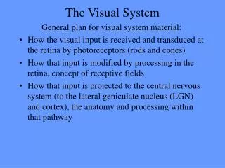
The Visual System May 30, 2014. Unit Two – Sensation & Perception. How do we interact with our environment? How do we process & interpret this information? Sensation Perception. Processing Visual Information. Why Vision?. Accurate representation of the world Respond to changes .

The Visual System May 30, 2014
E N D
Presentation Transcript
Unit Two – Sensation & Perception How do we interact with our environment? How do we process & interpret this information? Sensation Perception
Why Vision? Accurate representation of the world Respond to changes
Seeing is Believing What you see based on perception • Product of brain, biology Experience of the environment • Reality? • “Rebuilt” from components First, the anatomy…
The Eye Sensory receptors in the eye Signal from the environment is light • Response to light patterns • Iris is contractile tissue – constrict/dilate to change light entry through pupil
The Eye • lens – bends (refracts) light • Held/modulated by ciliary muscles • Aided by cornea • Depends on distance • Focuses light on retina– at back of eye • Image inverted • Near/far sightedness
The Retina Receptors for vision
Rods & Cones (20:1) Cones High acuity (details) Daylight Photopigments Rods Low acuity Night/low light vision (high sensitivity) No color pigments
Rods & Cones Fovea – center of retina w/ high acuity (cones) Little to no cones in periphery (almost all rods) • What can we see in center vs. periphery?
Acuity/Sensitivity Constant saccades & drifts – scan, never fixed Summation of constantly changing info - fovea
Acuity/Sensitivity • Constriction - focus of light on fovea • Acuity, details • Dilation - diffuse light to fovea & periphery • Sensitivity, dimly lit • Low light - sacrifice acuity for sensitivity • SNS - pupil dilation – does this make sense?
Retina Receptors bipolar cells ganglion cells
The Retina Acuity is a property of low convergence
Receptor Action No action potentials… • Rhodopsin (pigment) absorbs light • G-protein coupled receptor • Depolarization/hyperpolarization of cell • Release of neurotransmitter (Diagram in text)
Receptive Fields • Respond to some aspect of stimuli • Neurons work together – info about stimuli • Position in space, orientation, boundaries, movement
Color Vision • Cones - pigments respond to different wavelengths • Wavelength determines color • Humans – “red, green, blue” • Color - combination of activation of these receptors • Proportion of activity in different cones
Color Vision Wavelength - color perception Intensity – brightness Color constancy – relative, indep. of lightsource
Color Vision Not the light itself that is colored or bright… …our perception/ interpretation
Color Vision • Color blindness - cannot distinguish wavelengths • Two photoreceptors/pigments do not differ • Usually red/green
Retina Cortex Receptors ganglion cells Leave eye (optic nerve) for processing Blind spot (no receptors) • Masked w/ both eyes • System “fills in”
Retina Cortex • To visual cortex (perception) • Via the thalamus (retina-geniculate-striate pathway) • Also to superior colliculi in midbrain – why? • Sleep/wake • Eye movements/attention
Retina Cortex • Visual field – corresp. to particular spot on retina • Nasal, temporal hemiretina • Info from contralateral visual field • Cross over- optic chiasm • Optic nerve (L/R eye); Optic tract (L/R visual field) • Effect of damage depends on location
Retina Cortex • Info (shape, color, etc) to thalamus • Parvocellular cells – small, fine detail (cones) • Magnocellular cells – large, movement (rods) • To cortex for further processing Different aspects of stimulus processed separately! Still highly organized
Area V1 • Primary visual cortex - posterior occipital cortex • Topographic organization (conserves relationship) • Adjacent retina adjacent in LGN & cortex • Fovea periphery : posterior anterior • Lower visual field in upper region (& v. versa)
Area V1 • Visual experience – maintain, fine tune connections • Sensitive period • Plasticity • Representation of stimulation as “visual” • What would happen if V1 stimulated by auditory nerve?
Binocular Vision Why do we have two eyes? • See in almost every direction without turning head • Allows 3-D vision (depth) • Overlap (both eyes at the same time)
Binocular Vision • Disparity – diff. betw. where info falls on each retina • Cue for distance • Cells in cortex sensitive to disparity • Strabismus alters normal binocular vision
Binocular Vision • Predators – eyes in front, more overlap • Can also have more fovea • Better depth perception • Prey – eyes on sides, sacrifice depth • Judge distance of prey, acuity • Widen range of vision, predators approaching
Vision Across Species http://i.imgur.com/aRjn8.png
Review Light = stimulus, receptors on retina Retinal receptors thalamus (& brainstem) primary visual cortex Primary cortex – orientation, edges (receptive field) Higher order processing
A Holistic View See things as whole rather than a collection of parts… …but each area of cortex does not Each has unique “perspective” - require coordination between these regions How does the brain see objects as a whole?
Beyond Area V1 • Reassemble features into objects • Primary cortex to secondary sites • Specialization • Area V4 for color perception
Area V5/MT • Motion • Info from primary cortex & magnocellular cells • Form, speed, direction and location • Poor acuity
Area V5/MT • Motion parallax – if cross retina quicker, appear to move quicker • Cue for depth • Akinetopsia – difficulty w/ movement progression • Series of stills
Area V5/MT • Responding to movement across the retina.. … so what is the problem? • Did object or eye move? • Internal sense
Parallel Processing Secondary Association cortex Dorsal stream • Ego (person) centered • Where it is? • How to interact w/ it
Parallel Processing Ventral stream Object centered What is it? Work together to guide interaction - w/ other sensory info
Dorsal Stream • (“Where”) … or how: behavioral interactions • Spatial relationship • Representation in space -- grasping, manipulations