Download
1 / 30
300 likes | 369 Vues
Understand bronchiolitis, croup, asthma, pneumonia, and other respiratory conditions in children, their symptoms, and physiotherapy management. Learn about risk factors, pathophysiology, and treatment options for common childhood lung ailments.

E N D
Respiratory diseases in Childhood Robyn Smith Department of Physiotherapy UFS 2011
Bronchiolitis • Seasonal disease, and is common in winter months • Most commonly caused (60% cases) by RSV (Respiratory Syncitial Virus) • Most common severe lower respiratory tract infection in infancy • Mainly affects infants <2 years
Bronchiolitis Pathophysiology: • Viral infection causing inflammation of the bronchioles. • This leads to necrosis and destruction of the cilia and epithelial cells • Leads to obstruction of the small airways
Bronchiolitis Increased risk • Prematurity • Immuno-compromised children e.g. HIV infected infants • Chronic lung and heart diseases
Bronchiolitis Clinical signs and symptoms • Initially looks like a common cold • Develops a dry cough and difficulty feeding • Wheezing • Respiratory distress
Bronchiolitis • Management consists of Oxygen therapy • Minimal handling • CPT not indicated especially if wheezing still present • May require intubation or trachae • Only if secondary infection develops • Tenacious nasal secretions might need clearing
Croup/laryngotracheobronchietis • = laryngeal infection • Child has a hoarse barking cough and stridor • No indication for CPT • Oxygen therapy • Adrenaline inhalations • Minimal handling • Usually clears spontaneously within 12-48 hours • May require intubation (CIP) or tracheostomy • CPT and suctioning may make it worse. May however suction in the presence of an artificial airway
Asthma • No universally accepted definition • Asthma is a lung disease with the following characteristics: • Reversible airway obstruction either spontaneously or with treatment • Airway inflammation • Increased airway responsiveness to a variety of stimuli
Asthma • No active CPT is child is wheezing or has a silent chest • CPT can exacerbate bronchospasm • Inhalation therapy • Dyspnoea management • May be indicated if child is ventilated or a secondary lung infection developed
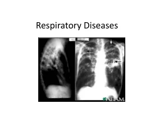
Lobar Pneumonia • Infection with consolidation of one or more lobes • Pleuritic pain common • CPT only indicated once the pneumonia is in resolution and the child is productive • Postural drainage • Active CPT including: Postural drainage Manual techniques e.g. percussions, vibration, shaking Breathing exercises Inhalation therapy if indicated
Bronchopneumonia • Acute inflammation of the bronchi and bronchioles with collapse and consolidation of associated groups of alveoli • Scattered irregularly throughout the lung • More often in lower lobes • No consolidation so one can immediately commence with CPT • Active CPT including: • Postural drainage • Manual techniques e.g. percussions, vibration, shaking • Breathing exercises • Inhalation therapy if indicated
Pnemocystis jiroveci (PCP) • Pneumocystis jiroveci pneumonia (formerly called Pneumocystis carinii or PCP) is most common opportunistic infection found in HIV positive patients • Patients are often acutely ill on admission with severe respiratory distress leading to respiratory failure failure requiring ventilation • Often do poorly despite maximal ventilation • Unstable • Subsequently many of them develop ARDS • Have an oxygenation and not a ventilation problem (thickening of the respiratory membrane with impaired gaseous exchange) • Minimal white frothy secretions and unproductive cough
Pnemocystis jiroveci (PCP) Physiotherapy: • Unstable – sensitive to position changing • Minimal handling • Often sound clear on auscultation, minimal secretions • Postural drainage if indicated • Active CPT if indicated • Proning to improve V/Q mismatch
Pertussis (whooping cough) • Necrosis of surface epithelium of the respiratory tract, which becomes covered in thick purulent exudate. This blocks the bronchi and bronchioles causing atelectasis. • Paroxysmal coughing spells • Child becomes cyanosed and red in the face • CPT not indicated during the acute stage • If atelectasis and mucous plugs are present may become indicated
Foreign body aspiration • CPT is only indicated post bronchioscopic removal of the foreign body. • Usually to treat underlying collapse or atelectasis
Bronchiectasis • Chronic inflammation of the bronchi with destruction of the cilia. • Resulting in impaired drainage of secretions leading to persistent lung infections of affected segments and lobes • Commonly associated with CF, pertussis andimmunodeficiency (HIV) • Child has a productive cough with excessive, purulent secretions
Bronchiectasis • Active CPT during exacerbation • Essential to teach a home clearance programme is taught including Postural drainage forced expiratory techniques Inhalation therapy • Breathing and thoracic mobility exercises • Activity to improve exercise tolerance
Cystic Fibrosis • Hereditary disorder of the exocrine glands and is characterised by hypertrophy and hyperplasia of the mucus secreting glands • CPT is important to assist in the clearance of secretions through
Cystic Fibrosis • Postural drainage routine and home programme • Active CPT • Inhalation therapies • IPPB • Active cycle of breathing • Forced expiration techniques • ↑physical activity • Breathing exercises • Trunk mobility and postural correction
Pulmonary Tuberculosis • Exposure to Mycobacterium Tuberculosis • Deposits in the lung and causes a primary infection • Physiotherapy: Breathing exercises Manual CPT techniques for areas collapse postural drainage if associated bronchiectasis mobilization
Lung tumours • Controlled breathing exercises • Gentle vibrations. Vigorous percussions, vibrations and shaking are contra-indicated due to the poor general condition of the patient, possibility of haemoptysis, and presence of metastases of the underlying ribs or spine • Postural drainage
A lot of patients we see in the PICU have trauma related injuries. Common trauma related injuries include: Pedestrian or motor vehicle accidents Falls from a height Gunshot wounds Knife wounds Assault and physical abuse cases Trauma related injuries
Trauma Pneumothorax • Accumulation of air or gas in the pleural cavity • Compressing the lung • Can occur spontaneously or due to trauma • No CPT is to be performed before the pneumothorax is drained by inserting a intercostal drain • Positioning • Older children mobilization, breathing exercises, coughing with drain support and shoulder girdle exercises are important
Trauma Lung contusion • No active CPT if there is still active bleeding • CPT helps in improving lung expansion
Trauma Rib fractures and flail chest • Patient should be given adequate analgesia • May need ventilation and PEEP • Breathing exercises • Assisted coughing by stabilizing cheat wall with hands may be indicated • Use of mechanical vibrations above percussion??? • No shaking and manual vibration • Positioning and postural drainage as injuries allow
References • Images courtesy of GOOGLE • Paediatric dictate (2009) • Downie, P. A. 1992. Cash’s Textbook of chest, heart and vascular disorders for physiotherapists. 4 ed. • Poutney, T. 2007. Physiotherapy for children • Morrow, B. Chest physiotherapy in PICU. Red Cross children’s Hospital, UCT