ENZYMES
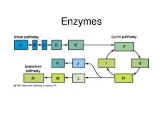
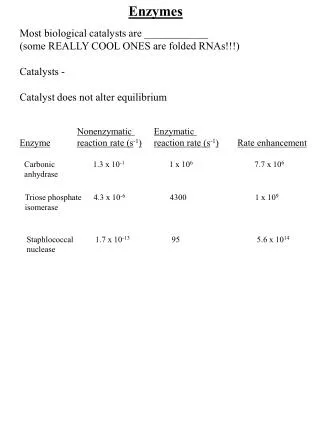
ENZYMES. Proteins that function as biological catalysts RNA catalysts are called ribozymes. S ↔ P . S = substrate(s) or reactant(s) & P = product(s). Enzyme Names S + type of reaction + ‘ ase ’ However, there are numerous exceptions.

ENZYMES
E N D
Presentation Transcript
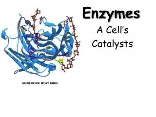
ENZYMES Proteins that function as biological catalysts RNA catalysts are called ribozymes S ↔ P S = substrate(s) or reactant(s) & P = product(s) Enzyme Names S + type of reaction + ‘ase’ However, there are numerous exceptions Normal – hexokinase, lactate dehydrogenase Abnormal – trypsin, DNA polymerase
catecholoxidase (1.10.3.1); pyruvatedehydrogenase (1.2.4.1) • hexokinase(EC 2.7.1.1); pyruvatekinase (2.7.1.40); RNA Polymerase (2.7.7.6) 3. carboxypeptidaseA (3.4.17.1); acetylcholinesterase (3.1.1.7) 4. carbonic anhydrase (4.2.1.1); adenylatecyclase (4.6.1.1) 5. phosphoglucoseisomerase (5.3.1.9); phosphoglucomutase (5.4.2.2) 6. DNA Ligase (6.5.1.1); acetyl CoA synthetase (6.2.1.1)
Assignment Look up in Swiss Prot – UniprotKB database and Wikipedia the following enzymes (human) 1) lactate dehydrogenase 4) carbonic anhydrase 2) glucokinase 5) phosphoglucomutase 3) carboxypeptidase A 6) DNA Ligase Write …… Name of enzyme & EC# equation for reaction Statement of its purpose/function of enzyme http://www.expasy.org/
S P G = G+ RT ln ([P]/[S]) G = G when all [ ]s = 1M (except [H+] = 1 x 10⁻⁷M pH = 7.0) At equilibrium G = 0 and DGº' = -RT ln ([P]/[S]) G‡(activation energy) determines the rate of the reaction An Enzymeincreases the rate at which a reaction achieves equilibrium by lowering the activation energy, Ea. It does not alter DGº′ nor change the final equilibrium state.
G = G+ RT ln Q where Q = ([P]/[S]) for the reaction S → P A reaction is proceeding in a cell. Which of the following best describes the free energy for this reaction. a) DG is (–) and constant. b) DG°′ must be (–). c) DG is (–) and increasing. d) None of the above
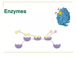
ES# ↔ EP MichaelisMenten Model k1 k2/kcat E + S ES E + P k-1 k-2 1 Enzyme (E) bind to substrate (S) Lock & Key model The Enzyme active site has a complementary shape to S Weak bonds are formed between E & S H-bonds, salt bridges, hydrophobic interactions & … occasionally covalent bonds
Is shape the only factor that determines bonding between E and S? a) yes b) no Lock & Key Model E & S have complementary shape E & S form complementary interactions – H-bonds, salt bridges, and/or hydrophobic interactions
Enzyme-Substrate Complex PDB 7GCH
Enzymatic Substrate Selectivity No binding Binding but no reaction Example: Phenylalanine hydroxylase
ES# ↔ EP MichaelisMenten Model k1 k2/kcat E + S ES E + P k-1 k-2 2 Enzyme converts S into P The enzyme converts S into a conformation that looks like S#. This requires some bonds in S to be weakened/stretched. The enzyme positions multiple substrates so that new bonds Can form when the ‘weakened’ bond(s) in S# breaks.
Initial rate assumption: [P] = 0 so k-2 [E] [P] = 0 Michaelis Menten Model k1 k2 E + S ES E + P k-1 k-2 0 rate of ES formation = k1 • [E] • [S] + k-2 • [E] • [P] rate of ES dissappearance = k-1 • [ES] + k2[ES] = (k-1 + k2) [ES] Steady state assumption – [ES] is constant = k1 • [E] • [S] = k-1 [ES] + k2 [ES] = (k-1 + k2) [ES] v = Vmax [S] (KM + [S]) KM = (k-1 + k2)/k1
Michaelis Menten Model k1 k2 E + S ES E + P k-1 k-2 v vs. [S] plot is hyperbolic 1/v = KM/Vmax•1/[S] + 1/Vmax Vmax = 1/yint & KM = slope • Vmax = -1/xint v = Vmax [S] (KM + [S])
Vmax KM Carbonic Anhydrase v vs. [S] HCO3- + H+→ CO2 + H2O v Experiment: Set up reactions with constant [E], and varying [S] Measure reaction rate (v) for each [S] Plot v vs. [S] and 1/v vs. 1/[S] [E] = 1 x 10⁻⁶ M Vmax = 1 Ms-1 KM = 0.012 M kcat = 1 x 10⁶ s-1 [S]
1/v = KM/Vmax * 1/[S] + 1/Vmax Double Reciprocal Plot Carbonic Anhydrase KM = -1/Xint = slope•Vmax 1/v slope = KM/Vmax Yint = 1/Vmax Xint = -1/KM 1/[S]
KM is ... (k-1 + k2)/k1 - combined rate constant [S] at which v = Vmax/2 KM = tighter binding Vmax (M•s-1) is .... v at Enzyme saturation: [ES] = [E]tot = k2 • [E]tot k2 (or kcat)called turnover # (s-1) independent of [E] An efficient enzyme will have ….. ? a) a large value for KM b) a large value for kcat c) both Enzyme efficiency [S] >> KM kcat Diffusion limited if kcat/KM ~ 1 x10⁸ to 1 x 10⁹ [S] << KM kcat/KM
Enzyme efficiency is limited by diffusion:kcat/KM • Can gain efficiency by having high velocity or affinity for substrate • Catalase vs. acetylcholinesterase
ENZYME INHIBITORS Competitive Noncompetitive I binds to active site I binds elsewhere I prevents ES E+P I prevents S binding KM - Vmax same Vmax - KM same I structurally similar to S I not similar to S
Vmax is represented by which Letter? Which letter represents KM?
Vmax is represented by which of the following? a) Yint b) slope c) 1/Yint d) slope/KM
How does Inhibitor type affect ….. • Reaction process • v vs. [S] plot • 1/v vs. 1/[S] plot • KM and Vmax.
competitive uncompetitive noncompetitive
Dihydrofolic acid Folic acid An important vitamin in humans, dihydrofolate is synthesized from Para-aminobenzoic acid (PABA) in bacteria. RDI = 400 mg/day. (1000 in pregnant women). Sources: Green leafy vegetables, fruits, nuts, beans, peas, dairy. (particularly high in asparagus and Brussel sprouts.)
H2N- -COOH BACTERIA PATHWAY GTP folK folQ folE folC folP dihydrofolate PABA DHPS DHFS Glutamate dihydropteroate PABA
Sulfa Drugs NH2 Icomp S = PABA NH2 C=O | OH NH2 O=S=O | NH2 O=S=O N-H N S sulfathiazole
Sulfthiazole resistance case study 1985 – 5 isolates of resistant StreptoccoccusPyogenessavedfrom patients in Sweden Hospital 1990’s – Genomes from normal and resistant isolates compared – highly mutated genes cloned & expressed in E. coli. DHPS gene found to be mutated. KMKi DHPS Kinetics G1 (suscep)0.7mM 0.2mM G56 (res) 2.5mM 27.4mM Difference 3.6x 137x Which enzyme will have better evolutionary ‘fitness’ in the absence of Inhibitor? a) susceptible b) resistant The resistant enzyme is less viable in absence of inhibitor but natural selection favors resistant form in presence of sulfa drugs.
Today’s topics: Review Enzyme Inhibitor type Carboxypeptidase Mechanism Enzyme Regulation What type if inhibitor is represented here? a) competitive b) noncompetitive c) uncompetitive Which of the following best describes this inhibitor? a) It must have a similar structure to the substrate b) It prevents the substrate from binding c) It increases Vmax. d) None of the above
I is structurally similar to S binds active site Prevents E + S ↔ ES ↑ KM Vmax same I binds to a separate site prevents ES → E + P ↓Vmax KM same I binds neat active site if S is already bound then prevents ES → E + P if S is not bound then prevents E + S → ES ↓Vmax KMcould go up or down depending …
Tryptathionone (oxidized) + NADPH ↔ Tryptathionone(reduced) + NADP+ Noncompetitive Inhibition
Tryptathionone (oxidized) + NADH ↔ Tryptathionone(reduced) + NAD+ Noncompetitive Inhibition Fig. 1: lineweaver-Burk plots showing the simple linear non-competitive inhibition of trypanothionereductase by TNQ2 either to NADPH (left, [trypanothione] constant at 100 µM) and trypanothione (right, [NADPH] constant at 200 µM); open circles, [TNQ2] = 0; closed circles, [TNQ2] = 1 µM; open squares, [TNQ2] = 2.5 µM; and closed squares, [TNQ2] = 5 µM
Alcohol Dehydrogenase un-competitive inhibition NAD + alcohol ↔ aldehyde/ketone + NADH Fig. 2. Inhibition of human ADH1C*2 by 5α-androstan-17β-ol-3-one (5α-dihydrotestosterone). The enzyme was assayed at 37 °C in 83 mM potassium phosphate, pH 7.3, 40 mMKCl and 0.25 mM EDTA buffer using a DU-7 spectrophotometer to measure the change in absorbance at 340 nm due to changing NADH concentrations (v=ΔA340 min−1). Duplicate assays were averaged. (A) The concentrations of 5α-androstan-17β-ol-3-one were 0 (●), 34 (■), 68 (▴) and 103 μM (♦) and the concentration of NAD+ was 0.5 mM. The data were fitted to the equation for uncompetitive inhibition, v=VA/(Km+A(1+I/Ki)), where V is the maximum velocity and A is the varied substrate. (B) The concentrations of 5α-androstan-17β-ol-3-one were 0 (●), 5 (■), 10 (▴) and 20 μM (♦) and the concentration of NADH was 0.1 mM. The data were fitted to the equation for competitive inhibition, v=VA/(Km(1+I/Ki)+A). The S.E. of the fits were <10%.
CARBOXYPEPTIDASE A (CPA) Digestive Enzyme: Zn Peptidase Family ― 309 aa’s―MW = 34,760 (Polypeptide)n→ (polypeptide)n-1 + C-terminal amino acid Example of Induced Fit X-ray Structure w/wo Substrate Mechanism Hypothesis How does the substrate bind to the active site? (KM effects) What features of this interaction contribute to the ‘turnover’? (Vmax effects)
H H | | H3N+- C - C - N-CH-CH2- -OH | || | H O COO- ARTIFICIAL SUBSTRATE = Gly-Tyr CPA – Substrate Comparison
Atom Residue Position relative to Fe C ARG127 -.01 C (CN3) ARG127+.43 C (CN3) ARG145+.50 C TYR248 +.50 OH TYR248+11.21 C GLU270 -.01 C (COO-) GLU270-1.64
Zn Gly-Tyr
Zn Gly-Tyr
Zn Substrate Y248
Y248-OH HOH HOH HOH HOH HOH HOH HOH E270 Zn +R127 +R145 H196 E72 H69
Y248 OH H H - CH-C-N-CH-CH2- O -OH O COO- E270 Zn +R127 +R145 H196 E72 H69 bonds contributing to KM & binding step.
Y248 Y248 OH OH H H -CH-C-N-CH-CH2- O -OH O COO- E270 Zn +R127 +R145 H196 E72 H69 H-OH
Y248 OH H H -CH-C-N-CH-CH2- O -OH O COO- E270 Zn +R127 +R145 H196 E72 H69 H-OH
+ H3N E270 The amino end KM but also Vmax since H2O substrate not in position. Y248 OH H H CH-C-N-CH-CH2- O -OH O COO- Zn +R127 +R145 H196 E72 H69
CARBOXYPEPTIDASE A (CPA) (Polypeptide)n→ (polypeptide)n-1 + C-terminal amino acid • CPA binds to S (a polypeptide) at the carboxy terminal end. • It prefers an aromatic or large nonpolar side chain. Binds to E NP pocket • The Zn coordinates to the C=O of S peptide bond (to be cut) • The carboxyl end of the molecule forms a salt bridge with Arg from E • Y248 – form H-bond with N-H of S peptide bond (to be cut) • The other substrate (H2O) forms 5th coordination site to Zn from E. • This is blocked by amino terminal end if S = dipeptide GY. • That is why GY is a poor substrate with tight binding (low KM) but low kcat.
O HHH || | | | - O - C- N- C - C - N-CH-CH2- | || | H O COO- H H | | H3N+ - C - C - N-CH-CH2- -OH | || | H O COO-
H H | | H3N+ - C - C - N-CH-CH2- -OH | || | H O COO- In Glycyl-tyrosine the + amino terminal end of the dipeptide … a) Forms a salt bridge with and Arg side chain from CPA. b) Is the 5th ligand to the Zn2+ of CPA. c) both of the above d) none of the above As a result of the interaction above Gly-Tyr has ….. a) A very small KM value. b) A very small Vmax value. c) both of the above d) none of the above
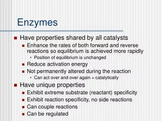
An enzyme is …. a) a protein b) a catalyst c) both of the above d) none of the above An enzyme will …. a) increase the rate of a reaction b) Increase the ratio of product to reactant at equilibrium c) both of the above d) none of the above