Download
1 / 81
820 likes | 920 Vues
This informative guide explores the differential diagnosis and management of cystic swellings in the neck, covering midline, lateral, and posterior regions. Learn about common cystic swellings like thyroglossal cysts, branchial cysts, and more, along with their embryology, pathology, clinical features, investigations, and treatments. Enhance your understanding of various neck masses and their complexities.

E N D
DIFFERENTIAL DIAGNOSIS OF CYSTIC SWELLINGS OF NECK AND MANAGEMENT By CIGIA PAUL
Swellings of neck • Midline • Lateral
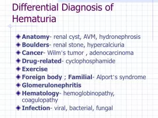
MIDLINE • Ludwig's angina • Enlarged sub mental lymph node • Sublingual dermoid • Sub hyoid bursitis • Pyramidal lobe • Pretracheal,paratrachael lymph nodes • Lipoma in suprasternal space of burns • Retrosternal goitre
Lateral swellings The Submandibular Triangle Enlarged lymph nodes Plunging ranula
The Carotid Triangle • Branchial cyst • Larngocoel • Cold abscess • Aneurysm of carotid artery • Carotid body tumour • Neurofibroma • Sternomastiod tumour
Posterior Triangle of Neck • Lymphangiomas • Cold abscess • Pharyngeal pouch • Heamangiomas • Cervical rib • Secondary nodes • Lipoma • Subclavian aneurysm
Common cystic swellings are • Thyroglossal cyst • Branchial cyst • Cystic hygroma • Dermoid cyst • Cold abscess • Plunging ranula • Pharyngeal pouch • Laryngocele • Sub hyoid bursitis
Thyroglossal cyst • Cystic swelling developed from remnants of thryoglossal duct Most common congenital neck mass:70% Second most common benign neck mass, after benign lymphadenopathy
The thyroid gland begins to develop in the 3rd week of fetallife as a median outgrowth from the floor of the primitive pharynx called Thyroglossal duct Distal bilobed ends lobes of thyroid This duct normally involutes by the 8th–10th gestational week. Embryology
Course of thyroglossal duct Passes from foramen caecum of the tongue between genioglossi muscles then it passes along midline downwards
Course of Thyroglossal tract It descends either in front of hyoid bone/ through the hyoid bone / hooks below and behind the hyoid bone Then descend downwards along midline to the upper border of thyroid cartilage and moves slightly to left and ultimately ending pyramidal lobe of thyroid gland
Fate of thryoglossal duct • Duct usually disappears except for the lower portion > thyroid gland • While the tract disappears ,a portion may remain patent which gives rise to cystic swelling due to accumulation of secretions in it > thyroglossal cyst
Pathology • Lined by stratified squamous epithelium or sometimes peudostratified ciliated columnar epithelium • Some lymphoid tissue outside epithelium: so prone to get infected
Pathology • Contents :- thick viscid mucus, • Cholesterol crystals may be present • Wall may contain thyroid tissue • Rarely : carcinomatous change occur(1%)
Clinical features Age : 15- 20 More incidence in women Presents with painless swelling in the midline of neck, Pain ,if infected No asso.systemic signs
On examination • POSITION • 1. Subhyoid • 2. At the level of thyroid cartilage • 3.Suprahyoid • 4.At the level ofCricoid cartilage • 5.Floor of mouth • 6. Beneath • Foramen caecum • skin normal unless infected
spherical /oval with long axis vertically along tract • Soft,cystc,fluctuant • Transilumination rarely positive • DON’T FORGET TO EXAMINE BASE OF TONGUE for ectopic thyroid • Mobility: 3 types • with deglutition • protrusion of tongue • horizontal
Complication • Recurrent infection • Fistula formation • Rarely papillary carcinoma
Investigations FNAC TFT USG ISOTOPE SCAN: for presence of normal thyroid tissue CT SCAN
Treatment • By surgical excision SISTRUNK PROCEDURE (Removal of cyst, the tract,central portion of hyoid bone,portion of tongue base upto foramen cecum) • Recurrence : less
Sistrunk procedure Before After
Branchial cyst Remnants of 2nd branchial arch
EMBRYOLOGY During third week of embryonic life, a series of mesodermal condensations (branchial arches) appear in the walls of primitive pharynx six in no,5th one disappears
EMBRYOLOGY 2 nd arch grows over 3rd and 4th Space between this is cervical sinus • Persistence of cervical sinus lead to brachial cyst • Inclusion of parotid epitheliun in upper deep cervical nodes
Pathology • Cysts are with fibrous wall lined by stratified squamous epithelium or sometimes peudostratified ciliated columnar epithelium • Wall contain plenty of Lymphoid tissue
Contains viscid, mucoid, cheesy material which contain cholesterol crystals in plenty
Clinical features • Age: 20-25 • 60% in males • Presents with a painless swelling in the upper and lateral part of neck • Become painful : if infected
On examination Seen on the upper and lateral aspect of neck, partly deep to strenocleidomastoid muscle 60%seen on left side 2% bilateral
Contd.. • round/oval of size, 5- 10 cm, • Smooth surface ,distinct edges, fluctuant • skin over swelling is normal • Translimination negative • Not freely mobile • Not reducible, compressible • On aspiration : cholesterol crystals
Complication • Recurrent infection • Brachial fistula formation
Investigations 1.FNAC : Contains viscid, mucoid, cheesy material which contain cholesterol crystals in plenty 2.USG 3.CT SCAN
Treatment Excision Don’t leave cyst Wall behind. Recurrence
Cystic hygroma Lymphangioma of neck A cystic swelling which contains multiple locules of clear lymph
Embryology • During embryonic life ,by sixth week lymph sacs develop: 6 in number • They are 1 pair jugular lymph sac 1 pair posterior cisterna chyli Retroperitoneal Sequestration of a portion of jugular sac
Pathology • Multiloculated lined by single layer of endothelium • Brilliantly transluminant • locules and cyst intercommunicate
Sites • Posterior triangle of neck • Cheek • Axilla • Groin • Mediastinum
Clinical features • Presents in Infancy with a lump lower third of neck • Size may vary • Round, smooth, indistinct margins,Soft cystic ,fluctuant • Impulse on cough • Partly compressible • Brilliantly translucent
Complication • Rapid growth cause respiratory difficulty • Infection
usg Investigation Fnac Usg C T Scan M R I
Treatment • Excision • Previously sclerosants advised: now CI • Newer therapy: Streptococcal product :OK432 • Recurrence >if incompletely removed
Sublingual dermoid cyst • Congenital sequestration dermoid
Pathology • Thin walled cyst • Lined by squamous Epithelium • Wall contains hair follicles, sweat glands, sebaceous glands • NEVER CONTAIN HAIR
TYPES • MIDLINE: inclusion of ectoderm in line of fusion of 1st branchial arch • LATERAL :These usually derive from 2nd branchial cleft
Supramylohyoid Inframylohyoid TYPES
Midline or to one side of floor of mouth Mucous membrane over is normal Smooth, definite edge, cystic swelling, fluctuation +, No transluminant Supramylohyoid
Inframylohyoid • Occupy submental region or submandibular region • Bimanually palpable • Fluctuation + • No transluminant • No movement with protrusion of tongue , deglutition
Investigations • F n a c • U s g • CT scan shows a well-defined mass in the submandibular space with a sack-of-marbles appearance • M R I
Treatment Excision Approach • Supramylohyoid : through mouth • Inframylohyoid: through neck
BEFORE AFTER
Plunging ranula • Ranula with cervical prolongation • Rana means frog