Download
1 / 150
1.53k likes | 1.97k Vues
Spinal Cord and Reflexes. Muse: Lecture #9 11/22/10. Spinal cord, nerves and reflexes. Figure 13–1 An Overview of Chapters 13 and 14. Central nervous system (CNS). Peripheral nervous system (PNS). Sensory (afferent) division. Motor (efferent) division. Somatic nervous system.

E N D
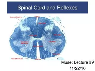
Spinal Cord and Reflexes Muse: Lecture #9 11/22/10
Spinal cord, nerves and reflexes Figure 13–1 An Overview of Chapters 13 and 14.
Central nervous system (CNS) Peripheral nervous system (PNS) Sensory (afferent) division Motor (efferent) division Somatic nervous system Autonomic nervous system (ANS) Sympathetic division Parasympathetic division accelerator brake Figure 13.1
Spinal Cord • Gross Anatomy of the Spinal Cord • About 18 inches (45 cm) long • 1/2 inch (14 mm) wide • Ends between vertebrae L1 and L2 • Bilateral symmetry • Grooves divide the spinal cord into left and right • Posterior median sulcus: on posterior side • Anterior median fissure: deeper groove on anterior side
Spinal Cord • Gross Anatomy of the Spinal Cord • The Distal End • Conus medullaris: • thin, conical spinal cord below lumbar enlargement • Filum terminale: • thin thread of fibrous tissue at end of conus medullaris • attaches to coccygeal ligament • Cauda equina: • nerve roots extending below conus medullaris
Spinal Cord Figure 13–2 Gross Anatomy of the Adult Spinal Cord.
Spinal Cord Figure 13–2 Gross Anatomy of the Adult Spinal Cord. Made by Honda?
Spinal Cord • 31 Spinal Cord Segments • Based on vertebrae where spinal nerves originate • Positions of spinal segment and vertebrae change with age • Cervical nerves: • are named for inferior vertebra • All other nerves: • are named for superior vertebra
Spinal Cord • Roots • Two branches of spinal nerves • Ventral root: • contains axons of motor neurons • Dorsal root: • contains axons of sensory neurons • Dorsal root ganglia • contain cell bodies of sensory neurons
Spinal Cord • The Spinal Nerve • Each side of spine • Dorsal and ventral roots join • To form a spinal nerve • Mixed Nerves • Carry both afferent (sensory) and efferent (motor) fibers
Spinal Cord Figure 13–3 The Spinal Cord and Spinal Meninges
Spinal Cord Figure 13–3 The Spinal Cord and Spinal Meninges
Spinal Cord • The Spinal Meninges • Specialized membranes isolate spinal cord from surroundings • Functions of the spinal meninges include • Protect spinal cord • Carry blood supply • Continuous with cranial meninges • Meningitis: • Viral or bacterial infection of meninges
Spinal Cord • The Three Meningeal Layers • Dura mater • Outer layer of spinal cord • Arachnoid mater • Middle meningeal layer • Pia mater • Inner meningeal layer
Spinal Cord • The Dura Mater • Tough and fibrous • Cranially • Fuses with periosteum of occipital bone • Is continuous with cranial dura mater • Caudally • Tapers to dense cord of collagen fibers • Joins filum terminale in coccygeal ligament • The Epidural Space • Between spinal dura mater and walls of vertebral canal • Contains loose connective and adipose tissue • Anesthetic injection site
Spinal Cord • The Arachnoid Mater • Middle meningeal layer • Arachnoid membrane • Simple squamous epithelia • Covers arachnoid mater
Spinal Cord • The Interlayer Spaces of Arachnoid Mater • Subdural space • Between arachnoid mater and dura mater • Subarachnoid space • Between arachnoid mater and pia mater • Contains collagen/elastin fiber network (arachnoid trabeculae) • Filled with cerebrospinal fluid (CSF) • Cerebrospinal Fluid (CSF) • Carries dissolved gases, nutrients, and wastes • Spinal tap: withdraws CSF
Spinal Cord • The Pia Mater • Is the innermost meningeal layer • Is a mesh of collagen and elastic fibers • Is bound to underlying neural tissue
Spinal Cord Figure 13–4 The Spinal Cord and Associated Structures
Gray Matter and White Matter • Sectional Anatomy of the Spinal Cord • White matter • Is superficial • Contains myelinated and unmyelinated axons • Gray matter • Surrounds central canal of spinal cord • Contains neuron cell bodies, neuroglia, unmyelinated axons • Has projections (gray horns)
Gray Matter and White Matter • Organization of Gray Matter • The gray horns • Posterior gray horns: contain somatic and visceral sensory nuclei • Anterior gray horns: contain somatic motor nuclei • Lateral gray horns: are in thoracic and lumbar segments; contain visceral motor nuclei • Gray commissures • Axons that cross from one side of cord to the other before reaching gray matter
Gray Matter and White Matter • Organization of Gray Matter • The cell bodies of neurons form functional groups called nuclei • Sensory nuclei: • dorsal (posterior) • connect to peripheral receptors • Motor nuclei: • ventral (anterior) • connect to peripheral effectors
Gray Matter and White Matter • Control and Location • Sensory or motor nucleus location within the gray matter determines which body part it controls
Gray Matter and White Matter • Organization of White Matter • Posterior white columns: lie between posterior gray horns and posterior median sulcus • Anterior white columns: lie between anterior gray horns and anterior median fissure • Anterior white commissure: area where axons cross from one side of spinal cord to the other • Lateral white columns: located on each side of spinal cord between anterior and posterior columns
Gray Matter and White Matter • Organization of White Matter • Tracts or fasciculi • In white columns • Bundles of axons • Relay same information in same direction • Ascending tracts: • carry information to brain • Descending tracts: • carry motor commands to spinal cord
Gray Matter and White Matter Figure 13–5a The Sectional Organization of the Spinal Cord.
Gray Matter and White Matter Figure 13–5b The Sectional Organization of the Spinal Cord.
Spinal Cord Summary • Spinal cord has a narrow central canal • Surrounded by gray matter • Containing sensory and motor nuclei • Sensory nuclei are dorsal SounD • Motor nuclei are ventral MoVe
Spinal Cord Summary • Gray matter • Is covered by a thick layer of white matter • White matter • Consists of ascending and descending axons • Organized in columns • Containing axon bundles with specific functions • Spinal cord is so highly organized • It is possible to predict results of injuries to specific areas
Spinal Nerves and Plexuses • Anatomy of Spinal Nerves • Every spinal cord segment • Is connected to a pair of spinal nerves • Every spinal nerve • Is surrounded by three connective tissue layers • That support structures and contain blood vessels
Spinal Nerves and Plexuses • Three Connective Tissue Layers of Spinal Nerves • Epineurium • Outer layer • Dense network of collagen fibers • Perineurium • Middle layer • Divides nerve into fascicles (axon bundles) • Endoneurium • Inner layer • Surrounds individual axons
Spinal Nerves and Plexuses Figure 13–6a A Peripheral Nerve.
Spinal Nerves and Plexuses • Peripheral Nerves • Interconnecting branches of spinal nerves • Surrounded by connective tissue sheaths
Spinal Nerves and Plexuses • Peripheral Distribution of Spinal Nerves • Sensory nerves • In addition to motor impulses: • dorsal, ventral, and white rami also carry sensory information • Dermatomes • Bilateral region of skin • Monitored by specific pair of spinal nerves
Spinal Nerves and Plexuses Figure 13–7b Peripheral Distribution of Spinal Nerves.
Spinal Nerves and Plexuses Figure 13–8 Dermatomes.
Spinal Nerves and Plexuses • Peripheral Neuropathy • Regional loss of sensory or motor function • Due to trauma or compression • chronic can be due to diabetes
Spinal Nerves and Plexuses • Nerve Plexuses • Complex, interwoven networks of nerve fibers • Formed from blended fibers of ventral rami of adjacent spinal nerves • Control skeletal muscles of the neck and limbs
Spinal Nerves and Plexuses • The Four Major Plexuses of Ventral Rami • Cervical plexus • Brachial plexus • Lumbar plexus • Sacral plexus
Spinal Nerves and Plexuses Figure 13–10 Peripheral Nerves and Nerve Plexuses.
Spinal Nerves and Plexuses Figure 13–10 Peripheral Nerves and Nerve Plexuses.
Spinal Nerves and Plexuses • The Cervical Plexus of the Ventral Rami • Includes ventral rami of spinal nerves C1–C5 • Innervates neck, thoracic cavity, diaphragmatic muscles • Major nerve • Phrenic nerve (controls diaphragm)
Spinal Nerves and Plexuses Figure 13–11 The Cervical Plexus.
Spinal Nerves and Plexuses Includes ventral rami of spinal nerves C5–T1 • The Brachial Plexus of the Ventral Rami • Major nerves of brachial plexus • Musculocutaneous nerve (lateral cord) • Median nerve (lateral and medial cords) • Ulnar nerve (medial cord) • Axillary nerve (posterior cord) • Radial nerve (posterior cord)
Spinal Nerves and Plexuses Figure 13–12a The Brachial Plexus.
Spinal Nerves and Plexuses Figure 13–12b The Brachial Plexus.
Spinal Nerves and Plexuses Figure 13–12c The Brachial Plexus.