Download
1 / 23
230 likes | 261 Vues
Explore a case of Kikuchi-Fujimoto disease involving CNS, characterized by lymphadenopathy, fever, and benign inflammation. Learn about histopathology, clinical features, differential diagnosis, and follow-up MRI findings.

E N D
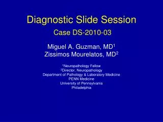
Diagnostic Slide Session Case DS-2010-03 Miguel A. Guzman, MD1 Zissimos Mourelatos, MD2 1Neuropathology Fellow 2Director, Neuropathology Department of Pathology & Laboratory Medicine PENN Medicine University of Pennsylvania Philadelphia
CLINICAL INFORMATION • 44 yo woman with headache, poor balance and MRI that revealed a right occipital lobe tumor. • Long standing history of intermittent lymphadenopathy: • 1981 (15 yo): Fever and left axillary lymphadenopathy. Biopsy “reactive/benign adenopathy” • 1991 (25 yo): Fever and cervical adenopathy: Biopsy: “reactive/benign” • 2007 (41 yo): Fever, night sweats with hypermetabolic retroperitoneal and mesenteric enlarged lymph nodes, clinically suspicious for lymphoma: Biopsy: “focally necrotizing lymphadenopathy, negative for malignancy or infection”. • The patient has history of allergies and multiple drug sensitivities with hypogammaglobulinemia detected in prior lab work up. Immunoglobulin levels now (performed on 1/19/09) show low IgA and IgM. • MRI shows an enhancing, intra-axial, right occipital lobe mass with surrounding T2 prolongation.
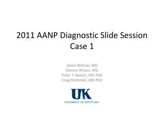
CD3 CD20 CD68 GFAP
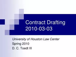
CD68 (Macrophages) CD8 (CytotoxicT cells) CD138 (Plasma cells)
KEY POINTS • Histopathology: • Nodular, Necrotizing inflammatory lesion with macrophages, lymphocytes (predominantly T-cells). • Absence of acute inflammatory component • Negative stains and cultures for micro-organisms • Clinical • Atypical MRI imaging mimicking tumor (differential included lymphoma or high grade glioma) • Long clinical history of previous diagnostic surgical procedure with negative histological findings for neoplasia in lymph nodes
Final Diagnosis Brain, right occipital, mass: Nodular, necrotizing, lymphohistiocytic inflammatory lesion consistent with CNS involvement by Kikuchi-Fujimoto disease.
Kikuchi-Fujimoto Disease (KFD)(Necrotizing histiocytic lymphadenitis without granulocytic infiltration) CLINICAL FEATURES Uncommon, self-limited, systemic lymphadenitis of unknown etiology Localized lymphadenopathy, predominantly in the cervical region, accompanied by fever and leukopenia in up to 50% of the cases. Generally benign and self-limited Sometimes: Recurrent lymphadenopathy Accompanying skin lesions Isolated fatal cases Rare associations: Following diffuse large B-cell lymphoma Stroma-rich Castleman's disease Lupus erythematosus Br J Radiol. 2003 Sep;76(909):656-8.
Kikuchi-Fujimoto Disease (KFD) • Pathogenesis • Necrosis due to cytoxic lymphocyte-mediated apoptotic cell death • Etiology unknown • Early suggestion that Toxoplasma may be involved not substantiated • Epstein–Barr virus (EBV), human herpesvirus (HHV)-6, HHV-8, and other viruses implicated: evidence not conclusive Source: Rosai: Surgical Pathology 9th edition
Kikuchi-Fujimoto Disease (KFD) • Histopathology • Paracortical necrotizing lesions • Focal • Well circumscribed • Abundant karyorrhectic debris • Scattered fibrin deposits • Collections of large mononuclear cells • Very scanty: plasma cells, neutrophils • Plasmacytoid monocytes: often numerous • if diffuse growth may simulate malignant lymphoma. • Occasionally: prominent secondary xanthomatous reaction • Electron microscopy: tubuloreticular structures and intracytoplasmic rodlets: similar to those in lupus erythematosus Source: Rosai: Surgical Pathology 9th edition Our case
Differential: Lymphoma (Hodgkin’s and non-Hodgkin’s) Infections including: -EBV/CMV -HIV -Cat Scratch Disease -Tuberculous adenitis -Autoimmune – in particular SLE is an important consideration as many patients initially diagnosed with Kikuchi’s dz have subsequently developed SLE (tubuloreticular structures in the lymphocytes and endothelial cells in SLE have been observed similar to those seen in Kikuchi’s dz) Kikuchi-Fujimoto Disease (KFD)
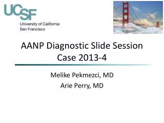
FOLLOW UP AFTER ONE YEAR Pre-operative MRI T1 post-gadolinium (1/19/2009) Follow up MRI T1 post-gadolinium (2/11/2010)
FOLLOW UP AFTER ONE YEAR Pre-operative MRI T1 post-gadolinium (1/19/2009) Follow up MRI T1 post-gadolinium (2/11/2010)
Comments • Involvement of Central Nervous System occasionally occurs in KFD typically in the form of aseptic meningitis • Brain parenchymal involvement by KFD is exceptionally rare, documented only by radiographic studies • In rare cases, KFD is associated with common variable immunodeficiency (of note is that this patient has hypogammaglobulinemia). References: • Hutchinson CB, Wang E. Kikuchi-Fujimoto disease. Arch Pathol Lab Med. 2010 Feb;134(2):289-93. • Chien YH, Yang YH, Hwu WL, Chou CC, Chiang BL. Common variable immunodeficiency with hypoglycemia, Kikuchi lymphadenitis, and hemiparesis in two siblings. J Microbiol Immunol Infect. 2003 Mar;36(1):65-8. • Shafqat S, Memon SB, Hyder S, Hasan SH, Smego RA Jr. Brainstem encephalitiswith Kikuchi-Fujimoto disease. J Coll Physicians Surg Pak. 2003 Nov;13(11):663-4.