Ch 16 Lateralization, Language & the Split Brain
Ch 16 Lateralization, Language & the Split Brain. Intro. Like most everything in our body, the brain is bilateral Left & right hemispheres are entirely separate except for the cerebral commisures connecting them Major differences exist between the functions of the hemispheres

Ch 16 Lateralization, Language & the Split Brain
E N D
Presentation Transcript
Intro • Like most everything in our body, the brain is bilateral • Left & right hemispheres are entirely separate except for the cerebral commisures connecting them • Major differences exist between the functions of the hemispheres • Lateralization of function • Split-brain patients: those whose hemispheres have been separated • Language is the most lateralized of all cognitive abilities • (Mostly left hemi) • Hemis have different abilities & can function independently
Cerebral Lateralization of Function • Broca’s area: • Inferior prefrontal cortex of the left hemisphere • Patients with aphasia (inability to produce or comprehend language) had damage to this area • Apraxia (difficulty performing movements when asked to do so out of context) almost always associated with left hemi damage, even though symptoms are bilateral
Cerebral Dominance • Cerebral dominance: Idea that one hemi (usually left) plays the dominant role in controlling all complex behavioral & cognitive processes • So the left hemi is commonly called the dominant hemisphere & the right is the minor hemisphere
Tests of Cerebral Lateralization • Sodium Amytal Test: • During neurosurgery, inject sodium amytal to anesthetize one hemisphere of the brain & have patient recite a series of words • When in left hemi, patient becomes mute for a few minutes • When in right hemi, no effect on language • Dichotic Listening Test: • Audio of #s being read played through headphones, with a different set of #s going to each ear (simultaneously) • When asked to repeat all the #s, most people say more #s heard in the right ear • Indicating left brain hemi for language; contralateral • Functional Brain Imaging: • PET or fMRI scans • During language tests, more activity is shown in the left hemi
Relation Between Speech Laterality & Handedness • Dextrals: right handers • Sinestrals: left handers • Study of handedness, hemisphere damage & aphasia showed that the left hemi is dominant for language for almost all dextrals & most sinestrals • Sinestrals are more variable in which hemi controls language
Split Brain • Corpus callosum: brain tissue that connects the 2 hemispheres • The largest cerebral commisure • Contains 200 million axons • A study using cats with transected (cut) corpus callosums showed that they were equally able to learn a task using one hemi as when using both • When tested using the opposite hemi, it was as if they had never learned it • Effectively showing the hemis acted as 2 separate brains • Conclusion: function of corpus callosum is to transmit info between the hemispheres
Commissurotomy in Human Epileptics • Commissurotomy: transecting the corpus callosum • Done as a treatment for severe epilepsy to prevent the spread of the over-stimulated signal from one hemi to the other • Tests done by delivering info to one hemi while keeping it out of the other • Like with split-brain animals, split-brain humans seem to have 2 independent brains, each with its own stream of consciousness, abilities, memories & emotions • Unlike the animals, human hemis are unequal in their abilities to perform certain tasks • Especially left hemi is capable of speech, right is not
Hemispheres Functioning Independently • Reminder: Input from one visual field or movement/feeling from one hand go to the contralateral hemisphere
Hemispheres Functioning Independently • Left hemisphere can tell what it has seen, right hemisphere can show it. • Studies of split-brain patients: • Present a picture to the right visual field (left brain) • Left hemisphere can tell you what it was • Right hand can show you, left hand can’t • Present a picture to the left visual field (right brain) • Subject will report that they do not know what it was • Left hand can show you what it was, right can’t
Doing 2 Things at Once • Your brain can learn 2 different things at once • When shown 2 different pictures (one in each visual field), patients can reach into 2 bags (one with each hand) and correctly grab the items they saw • However, if you ask them what was in their hands, they would say 2 of what was shown on the right & be surprised when they looked at the objects in their hands and saw 2 different items
Doing 2 Things at Once • Experiment is repeated, but instead of reaching into bags, the patient can see the objects in front of them • When the patient is asked to pick up what was seen sometimes the helping-hand phenomenon occurs • This is when the right hand goes to pick up what was seen by the left hemi & the right hemi “realizes” that is the wrong object (not what the right hemi saw) & causes the left hand to shoot out to redirect the right hand towards the correct object
Doing 2 Things at Once • Because the hemis are effectively seeing twice as much at once, split brain patients can find a visual target in a group of items more quickly than healthy individuals • Chimeric figures test • Visual completion • Scotoma (blind spot)
Split Brain Misc. • For most split brain patients, the left hemi tends to control most of everyday activities • However, in some cases, the right hemi has a will of its own & will create conflicts with the left hemi • Split brain hemis mostly act independently, but they can interact via brain stem • Individuals can vary on hemispheric independence • Emotional info about a picture presented to right hemi can be transferred to left hemi which can communicate the feeling, even when it doesn’t know what the picture was • More complex tasks tend to involve both hemis • Elderly display less lateralization of function
Differences between Left & Right Hemis • Many functions have no difference between the hemis • When there are differences, they tend to be a slight bias in favor of one hemi, NOT a clear cut, absolute difference • Functions do not reside exclusively in one hemi or the other • Language is the most lateralized cognitive ability, but even it is not totally absent from the right hemi • Right hemi language skills like that of a preschooler
Cerebral Lateralization of Function • Superiority of left hemi in controlling ipsilateral movement • Most movement controlled contralaterally, but some ipsilateral & left is better at it • Superiority of right hemi in spatial ability • Feeling an object in hand & deciding which 2-D image shows what it would look like unfolded • Specialization of right hemi for emotion • Better at identifying facial expressions of emotion • Superior musical ability of right hemi • Dichotic listening test with musical tunes, better able to identify with left ear
Cerebral Lateralization of Function • Hemispheric differences in memory • Both hemis involved in memory, but differ in which is best at certain tests • Left hemi specialized for episodic memory • Left hemi for memory of verbal info • Right hemi for nonverbal info • The hemis approach cognitive tasks in different ways • Left hemisphere acts as the interpreter; continuously assessing patterns of events and trying to make sense of them • Left hemi dominant for language, but right is better at perceiving intonation of speech & identifying the speaker • Example of how these functional lateralizations are not absolute
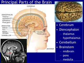
Anatomical Asymmetries of the Brain • Frontal operculum • In frontal lobe • In left hemi it is the location of Broca’s area • Planumtemporale • In temporal lobe; called Wernicke’s area • Involved in comprehension of language • Larger in left hemi, but only in 65% of brains • Heschl’sgyrus • In temporal lobe; primary auditory cortex • Larger in right hemi, often 2 gyri in right & only 1 in left • Difficult to define the exact border/size of these structures
Evolution of Cerebral Lateralization • Analytic-Synthetic Theory: • Left hemi operates in an analytical, logical, computerlike way; analyzing stimulus info input sequentially, collecting extracting relevant info & attaching a verbal label • Right hemi synthesizes; concerned with overall stimulus configuration and organizes & processes info in terms of wholes • Mostly pop psychology; difficult to test empirically
Evolution of Cerebral Lateralization • Motor Theory: • Left hemi is specialized for speech because it is a type of fine motor movements • Doesn’t explain why motor function would have become lateralized • Linguistic Theory: • Primary role of left hemi is language • Deaf people with left hemi damage have difficulty using sign language, but not pantomime
Evolution of Cerebral Lateralization • All classes of vertebrates have a right side preference for feeding • Once hands evolved (monkeys & apes), there was a right hand preference for feeding and other complex behaviors • Left hemi bias for communication in non-human species • Ex: birdsong, dogs & monkeys for calls of conspecifics
Advantages of Cerebral Lateralization • Advantageous for areas of the brain that perform similar functions to be located in the same hemi • May be more efficient for neurons performing a particular function to be concentrated in one hemi • 2 different kinds of cognitive processes can be more easily performed simultaneously if they are lateralized to diff hemis
Cortical Localization of Language • Language localization refers to the location with the hemis of the circuits that participate in language-related activities • Wernicke-Gerschwind Model • The predominant theory of language localization
Cortical Localization of Language • Broca’s area controls speech production • Wernicke’s area controls language comprehension • Broca’s aphasia: • Lesions of Broca’s area hypothesized to produce aphasia with symptoms that are primarily expressive • Normal comprehension of written/spoken language • Speech that retains meaningfulness despite being slow, labored, disjointed & poorly articulated) • Wernicke’s aphasia: • Lesions of Wernicke’s area produce aphasia with symptoms that are primarily receptive • Poor comprehension of both written/spoken language • Speech that is meaningless but still retains superficial structure, rhythm & normal intonation • Word salad
Cortical Localization of Language • Conduction aphasia: • Caused by damage to the pathway connecting Broca’s & Wernicke’s area (arcuate fasciculus) • Mostly intact comprehension & speech, with difficulty repeating words they just heard • Angular gyruscontrols comprehending language-related visual input • Area of left temporal & parietal cortex just posterior to Wernicke’s area • Damage to this area can cause alexia (inability to read) & agraphia (inability to write) • No difficulty speaking or understanding speech
Wernicke-Geschwind Model • 7 components (all in left hemi): • Primary visual cortex • Angular gyrus • Primary auditory cortex • Wernicke’s area • Arcuate fasciculus • Broca’s area • Primary motor cortex
Wernicke-Geschwind Model • During a conversation, auditory info is received by primary auditory cortex & sent to Wernicke’s area, where they are comprehended • To respond, Wernicke’s area generates the neural representation of the thought underlying the reply & transmits it to Broca’s area (via left arcuate fasciculus) where it activates the appropriate program of articulation that fires the neurons of primary motor cortex & then muscles of articulation
Wernicke-Geschwind Model • When reading aloud, signal received by primary visual cortex is transmitted to left angular gyrus, which translates visual form of word into its auditory code & transmits it to Wernicke’s area for comprehension • Wernicke’s area then triggers responses in arcuate fasciculus, Broca’s area & motor cortex to elicit speech sounds
Problems with the Wernicke-Geschwind Model • Many aspects of this theory are oversimplifications • Removal of Broca’s area but no surrounding area has no lasting effects on speech • Speech problems may be due to swelling of surrounding area • No permanent speech difficulties with lesions to arcuate fasciculus • No permanent alexia or agraphia with lesions of angular gyrus • Much of Wernicke’s area can be removed with no long term language deficits
Problems with the Wernicke-Geschwind Model • More recent data has shown: • No aphasic patients have damage restricted to just Broca’s or Wernicke’s area • Aphasic patients almost always have significant damage to subcortical white matter • Large anterior lesions are more likely to produce expressive symptoms; large posterior lesions more likely to produce receptive symptoms • Global aphasia (severe disruption of all language related abilities) is usually related to massive lesions of anterior cortex, posterior cortex & underlying white matter • Aphasic patients sometimes have brain damage not near the Wernicke-Geschwind areas
Further Study of the Wernicke-Geschwind Model • More detailed info about areas related to language function by testing with electrical stimulation (as opposed to damage/lesions) • Sites at which stimulation blocked/disrupted speech are scattered throughout frontal, temporal & parietal cortex (not just restricted to Wernicke-Geshwind areas) • No specific areas that caused specific speech disturbances (ex: pronunciation, naming objects) • Right hemi stimulation almost never disrupted speech • Major differences among individuals & their neural organization of language abilities
Current State of Wernicke-Geschwind Model • Original model supported in 2 ways • Broca’s & Wernicke’s areas play important roles in language • Tendency for aphasias associated with anterior damage to involve expressive deficits & posterior damage to involve receptive deficits
Current State of Wernicke-Geschwind Model • Original model not supported • Aphasia typically associated with widespread damage, not just to Wernicke-Geschwind areas • Aphasia can result even when damage does not involve any Wernicke-Geschwind areas • Broca’s & Wernicke’s aphasias rarely exist in pure forms; aphasia almost always involves both expressive & receptive symptoms • Major differences in locations of cortical language areas in different individuals • Generally, the theory has been abandoned by researchers
Cognitive Neuroscience of Language • Currently used in language research • Defined by 3 premises: • Premise 1: Each complex language related process (speech, comprehension, reading) is the combo of several constituent cognitive processes, which may be organized separately in different parts of the brain. • Premise 2: Areas of the brain involved in language are not dedicated solely to that purpose • Ex: language areas can also function in memory • Premise 3: Brain areas for language are small, widely distributed & specialized
Functional Brain Imagine & Localization of Language • fMRI study showed that areas of the brain involved in silent reading were patchy, variable among patients & not limited to classic Wernicke-Geschwind areas • Far more activity in left hemi • PET study of activity in temporal lobe while naming objects in categories • Involved brain areas outside classic Wernicke-Geschwindareas • Area activated depended on category of the objects
Cognitive Neuroscience of Dyslexia • Dyslexia: pathological difficulty in reading, not resulting from general visual, motor, or intellectual deficits • 2 types: • Developmental: • Becomes apparent as child learns to read • 5-11% of English speaking kids • 2-3x higher in boys than girls • Acquired: • Caused by brain damage in people who could already read • Rare
Developmental Dyslexia • Has a major genetic component (50% heritability) • No single kind of brain pathology has been found in all cases • Disorder has various forms, likely with different neural correlates • Results from disturbance of phonological processing (representation & comprehension of speech sounds)
Acquired Dyslexia • 2 types: • Surface: • Inability to pronounce words based on their memories of the words (lexical procedure) but can still apply rules of pronunciation in reading (phonetic procedure) • Difficulty pronouncing words that don’t follow common rules of pronunciation (ex: have, lose, steak) & will often incorrectly pronounce them based on rules (ex: rhyming with cave, hose, beak) • Deep: • Inability to apply rules of pronunciation in their reading (lost phonetic procedure) • Incapable of pronouncing nonwords