Case I
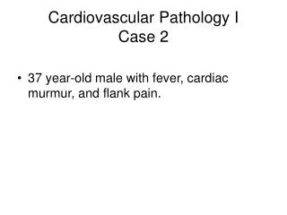
Case I. Patient Description . 41 year old, black male with history of HIV, HBV and Kaposi’s sarcoma presents with abdominal pain, fever and fatigue worsening over past month On efavirenz/tenofovir/emtricitabine, TMP-SMX, azithromycin, but on no KS therapy. Patient Description.


Case I
E N D
Presentation Transcript
Patient Description • 41 year old, black male with history of HIV, HBV and Kaposi’s sarcoma presents with abdominal pain, fever and fatigue worsening over past month • On efavirenz/tenofovir/emtricitabine, TMP-SMX, azithromycin, but on no KS therapy
Patient Description • Enlarged inguinal nodes bilaterally, palpable spleen, diffuse abdominal pain with some guarding, no palpable masses, active BS, no rectal masses • Few scattered KS lesion on lower extremities bilaterally, no edema • Remainder of exam normal
Patient Description • CD4+ count 56 cells/mm3; plasma HIV RNA 86 copies/mL • Liver enzymes mildly elevated, HBV quant 80c/ml, HCV quant neg, creatinine normal, amylase normal • Hb 9.7 g/dL; Hct 29.3%; WBC 3000/mm3; platelet count 120,000; MCV 90 fL; retic 0.014
Possible Diagnosis 1. GI Kaposi’s sarcoma with GI bleed 2. Non-Hodgkin’s lymphoma 3. Abdominal abscess, MAI or others 4. CMV colitis 5. Anal cancer 6. Progressive HIV/AIDS
Possible Diagnosis 1. GI Kaposi’s sarcoma with GI bleed 2. Non-Hodgkin’s lymphoma 3. Abdominal abscess, MAI or others 4. CMV colitis 5. Anal cancer 6. Progressive HIV/AIDS
Additional Tests 1. PET/CT abdomen 2. Biopsy lymph node 3. Repeat VL and HIV genotype 4. Culture blood and stool, endoscopy 5. Stool occult blood, bone marrow, further heme work up 6. Chest X-ray
Additional Tests 1. PET/CT abdomen 2. Biopsy lymph node 3. Repeat VL and HIV genotype 4. Culture blood and stool, endoscopy 5. Stool occult blood, bone marrow, further heme work up 6. Chest X-ray
Laboratory Findings • Urinalysis normal, bilirubin normal, urine hemosiderin negative, LDH 500 IU/L, G6PD level normal, ferritin level elevated. • Stool occult blood positive, blood cultures X 3 neg, MAC cultures neg, AFB negative, stool cultures neg, CXR normal • CT scan abdomen - ordered • HIV drug genotype - ordered • Bone marrow aspiration and biopsy- ordered
Laboratory Findings • CT scan of abdomen shows large gastric mass, enlarged perigastric and periaortic nodes, a few liver nodules and small amount of ascites • Gastroscopy shows large gastric ulcerated mass, no active bleeding • Biopsy reveals large B-cell NHL, CD-20+, no CMV or KS
Gastric lymphoma. Persistent gastric wall thickening (S). Enlarged spleen and an enlarged Lt. gastric lymph node (arrow)
Clinical Decision Point • The patient has no CNS symptoms, CBC has not changed. Staging work up includes: • Whole body PET/CT – hypermetabolic nodes in gastric, periaortic and mediastinal areas • LP with CSF cytology - neg for lymphoma • Bone marrow aspiration and biopsy • hypocellular, normal iron stores, no lymphoid infiltrates, normal lymphocyte flow panel, no granuloma or infection seen
Clinical Decision 1. Chemotherapy (e.g R-CHOP or R-EPOCH) 2. Hydrate, allopurinol, follow electrolytes, creatinine, phosphorus, calcium 3. Intrathecal cytosine arabinoside X 4 4. Continue TMP-SMX, azithromycin 5. Begin prophylaxis with antibacterial and antifungal medications 6. Follow CBC, +/- G-CSF 7. Change ART 8. All of the above
Clinical Decision 1. Chemotherapy (e.g CHOP-R or EPOCH-R) 2. Hydrate, allopurinol, follow electrolytes, creatinine, phosphorus, calcium 3. Intrathecal cytosine arabinoside X 4 4. Continue TMP-SMX, azithromycin 5. Begin prophylaxis with antibacterial and antifungal meds 6. Follow CBC, +/- G-CSF 7. Change ART 8. All of the above
Clinical Course • Patient tolerates chemotherapy with R-EPOCH, with apparent tumor response, however after two cycles, Hb now 7.0 g/dL. What do you do? 1. Repeat bone marrow aspirate and biopsy 2. Check stool occult blood, give iron and folic acid 3. Check EPO level and give recombinant erythropoietin alpha 4. Switch to new ARV 5. Transfuse 2 units of PRBC 6. Schedule endoscopy
Clinical Course • Patient tolerates chemotherapy with EPOCH-R, however after two cycles, Hb now 8.0 g/dL. What do you do? 1. Repeat bone marrow aspirate and biopsy 2. Give iron and folic acid 3. Check EPO level and give recombinant erythropoietin alpha 4. Switch to new ARV 5. Transfuse 2 units of PRBC 6. Schedule endoscopy • Transfused 2U PRBC with supplemental iron and folic acid • After three cycles of R-EPOCH, patient has achieved a complete remission. Treatment continues for 6 cycles. CBC returns to normal. • Patient followed q3 mos X 2 yrs the q 6 mo
Clinical Course • 5 yrs later, on routine exam he is found to have enlarged parotid and L neck nodes and mild anemia, Hb=11, CD4=210, VL<48 c/ml on darunavir/r and tenofovir/emtricitabine • What do you do? 1. Work up anemia, change ART, and follow nodes 2. Assume recurrent NHL and retreat with R-EPOCH, since relapse >12 mo post remission 3. Assume lymphoma and treat with alternate regimen 4. Assume progressive KS and treat with liposomal doxorubicin 5. Biopsy lymph node and bone marrow
Clinical Course • 5 yrs later, on routine exam he is found to have enlarged parotid and L neck nodes and mild anemia, Hb=11, CD4=210, VL<48 c/ml on darunavir/r and tenofovir/emtricitabine • What do you do? 1. Work up anemia, change ART, and follow nodes 2. Assume recurrent NHL and retreat with R-EPOCH, since relapse >12 mo p remission 3. Assume lymphoma and treat with alternate regimen 4. Assume progressive KS and treat with liposomal doxorubicin 5. Biopsy lymph node and bone marrow
Clinical Course • Biopsy of lymph node showed mixed cellularity Hodgkin lymphoma, CD15+, CD30+, CD20-, EBV+ • Patient treated with ABVD, G-CSF, prophylactic antibiotics and continued ART • Complete remission achieved after 3 cycles, patient treated for 6 cycles
Incidence of HL and NHL by CD4 count Biggar R J et al. Blood 2006;108:3786-3791
Distributions of CD4 counts 0 to 12 months before onset for Hodgkin lymphoma (HL) and non-Hodgkin lymphoma (NHL) n = 311 for HL cases; n = 4344 for NHL cases. Biggar R J et al. Blood 2006;108:3786-3791
Why the incidence of HIV-associated Hodgkin lymphoma may increase in the setting of improved immunity Gloghini and Carbone, Int J Cancer, 2007;120:2753-4
Clinical Course • 1.5 yrs later on routine follow up, again found to have enlarged left supraclavicular LN. Biopsy of lymph node showed mixed cellularity Hodgkin lymphoma, CD15+, CD30+, CD20-, EBV+ • PET/CT shows hypermetabolic LNs supraclav, partracheal, mesenteric, pericaval and periportal, BM and CSF negative for lymphoma • Patient treated with ICE, G-CSF, prophylactic antibiotics and continued ART and stem cells (CD34+) stored • Partial response noted after 2 cycles • What next?
Clinical Course • Because of high risk for recurrent or resistant disease, patient proceeds to autologous stem cell transplant. • BEAM (BCNU, etoposide, Ara-C, melphalan) conditioning given followed by autologous stem cells • ART continued • Prophylactic antibiotics and G-CSF given and LFTs monitored • Now 2 years after SCT patient in CR, back at work and continues to be closely followed.
Disease Free Survival after Autologous SCT Krishnan A. Biology of Blood and Marrow Transplantation 2010; 16:1302-1308
Overall Survival after Autologous SCT Krishnan A, et al Biology of Blood and Marrow Transplantation 2010; 16:1302-1308
Key Points • AIDS patients can have multiple cancers • Hogkins lymphoma may occur in patients with suppressed HIV and higher CD4 cts • Evaluate for multiple etiologies for anemia in advanced AIDS patients • Use prophylactic antibiotics and hematopoietic growth factors in AIDS patients on chemotherapy, especially if receiving rituximab or more myelo/immuno-suppressive regimens • Results of HDC-SCT in HIV have good long term outcomes