Optimising X-ray computer tomography images with a CT-simulator
220 likes | 388 Vues
Optimising X-ray computer tomography images with a CT-simulator. Philippe Van Marcke K.U.Leuven. Introduction. CT-simulator:. Introduction. computer program that immitates the working of a CT-scanner

Optimising X-ray computer tomography images with a CT-simulator
E N D
Presentation Transcript
Optimising X-ray computer tomography images with a CT-simulator Philippe Van Marcke K.U.Leuven
Introduction CT-simulator: Introduction • computer program that immitates the working of a CT-scanner • makes it possible to manipulate all parameters involved in the scanning process and investigate their effect • generating a lot of measurements in short time Development of the simulator Example of image optimisation Conclusions • useful tool for optimising image quality by trial and error
Defining objects Defining objects in the simulator: • superposing object parts • µ = µforeground - µbackground • different resolution for different parts Development of the simulator Development of the simulator Example of image optimisation Conclusions 1 2
Defining objects Defining objects in the simulator: • superposing object parts • µ = µforeground - µbackground • different resolution for different parts Development of the simulator Development of the simulator Example of image optimisation Conclusions 1 2 i
Defining objects Defining objects in the simulator: • superposing object parts • µp1 = µ1 • intensity of a monochromatic beam i passed through object part 1: Development of the simulator Development of the simulator Example of image optimisation Conclusions 1 i
Defining objects Defining objects in the simulator: • superposing object parts • µp2 = µ2- µ1 • intensity of a monochromatic beam i passed through object part 2: Development of the simulator Development of the simulator Example of image optimisation Conclusions 2 i
Simulator formula Intensity of a monochromatic beam i passed through an object consisting of P object parts: Development of the simulator Development of the simulator Example of image optimisation Conclusions • the simulator repeats this calculation a large number of times
Sampling the spectrum Monochromatic beams are grouped into polychromatic beams Development of the simulator Development of the simulator Example of image optimisation Spectrum is divided into K regions with an equal area Conclusions
Sampling the spectrum Monochromatic beams are grouped into polychromatic beams Development of the simulator Development of the simulator The formula for the monochromatic beam i: Example of image optimisation Conclusions
Sampling the spectrum Monochromatic beams are grouped into polychromatic beams Development of the simulator Development of the simulator The formula for the monochromatic beam i: Example of image optimisation Conclusions is extended to a polychromatic beam i:
Sampling source and detector Finite sizes of the source and detector elements are modelled by sampling both parts Development of the simulator Development of the simulator Example of image optimisation Conclusions source is modelled by S source samplesdetector elements are modelled by D detector samples
Sampling source and detector Finite sizes of the source and detector elements are modelled by sampling both parts Development of the simulator Development of the simulator The formula for the polychromatic beam i is repeated for all source and detector samples: Example of image optimisation Conclusions Implementing additional features: • noise • using hardware filters
Implementing noise and filters • Noise: Development of the simulator Development of the simulator Example of image optimisation fR number of frames used Conclusions • Filtration of the X-ray spectrum: µi attenuation coefficient of filter iti thickness of filter i
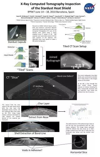
Image optimisation Simulating a dolomite sample (8 mm) with calcite veins Development of the simulator Example of image optimisation Example of image optimisation Conclusions (a) picture of sample (b) polychromatic simulation (1 frame) (c) polychromatic simulation (16 frames)
Image optimisation Simulating a dolomite sample (8 mm) with calcite veins Development of the simulator Example of image optimisation Example of image optimisation Conclusions (d) monochromatic simulation (e) polychromatic simulation (16 frames) using a 0,01 mm copper filter (f) polychromatic simulation (16 frames) using a 0,1 mm copper filter
Conclusions Conclusions: • Objects defined by superposing object parts • Polychromacity of the X-ray spectrum is modelled by averaging over a number of monochromatic simulations • The finite sizes of the source and detector elements are modelled by sampling both parts • It is possible to include noise and filters in the scanning process • Optimising image quality by trial and error experiments Development of the simulator Example of image optimisation Conclusions Conclusions