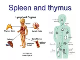
SPLEEN AND THYMUS
SPLEEN AND THYMUS. BRIG IQBAL MUHAMMAD KHAN MBBS, MCPS, FCPS ASSOC PROF OF PATHOLOGY HEAD OF HISTOPATH DEPT ARMY MEDICAL COLLEGE. SPLEEN. Normal measurements Structure Circulation Functions. STRUCTURE OF SPLEEN. Normal weight 150 g Invested by a thin connective tissue capsule

SPLEEN AND THYMUS
E N D
Presentation Transcript
SPLEEN AND THYMUS BRIG IQBAL MUHAMMAD KHAN MBBS, MCPS, FCPS ASSOC PROF OF PATHOLOGY HEAD OF HISTOPATH DEPT ARMY MEDICAL COLLEGE
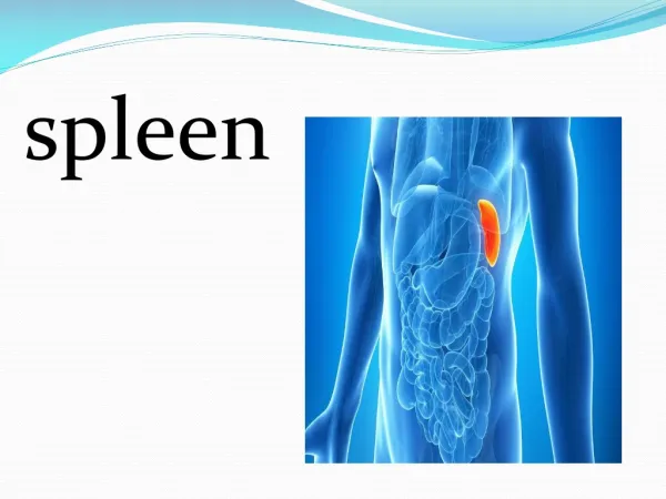
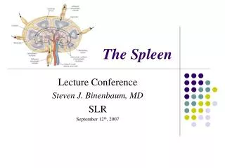
SPLEEN • Normal measurements • Structure • Circulation • Functions
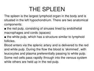
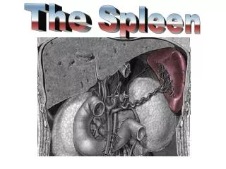
STRUCTURE OF SPLEEN • Normal weight 150 g • Invested by a thin connective tissue capsule • Cut surface show extensive red pulp dotted with white pulp • White pulp is composed of a central artery surrounded by a rim of T-lymphocytes, which at places expands to form lymphoid nodules, capable of developing germinal centres. • Red pulp is traversed by thin walled vascular sinusoids, separated by cords “Cords of Billroth”
STRUCTURE continue…… • The sinusoids are lined by discontinuous endothelium • The cords contain macrophages forming a labyrinth acting as filter • Circulation • Open • closed
FUNCTIONS OF SPLEEN • Phagocytosis of RBCs and Particulate matters • Antibody production • Haemotopoisis • Sequestration of formed blood elements SLENECTOMY result in increased susceptibility to sepsis to capsulated bacteria like pneumococcus, meningococcus and H. influenzae
CAUSES OF SPLENOMEGALY INFECTIONS • Parasitic : Malaria, Leishmaniasis, trypanosomiasis, schistosomiasis, echinococcosis, toxoplasmosis. • Bacterial : typhoid fever, T.B, brucellosis • Viral : Infectious mononucleosis, cytomegalovirus • Fungal : Histoplasmosis
CAUSES OF SPLENOMEGALY • LYMPHOHEMATOGENOUS DISORDERS Hodgkin’s disease, non Hodgkin’s lymphoma, leukaemia, multiple myeloma, myeloproliferative disorders, haemolytic anaemias, thrombocytopenic purpura • STORAGE DISORDERS Gaucher’s disease, Niemann – Pick disease, Mucopolysaccharidoses
CAUSES OF SPLENOMEGALY • CONGESTIVE STATES Cirrhosis of liver, portal or splenic vein thrombosis, Cardiac failure • IMMUNOLOGIC – inflammatory conditions Rheumatoid arthritis, SLE • MISCELLANEOUS Amyloidosis, primary and secondary neoplasms, cysts
MORPHOLOGY IN NONSPECIFIC ACUTE SPLENITIS • Mild to moderate splenomegaly (200-400 grams) • Acute congestion of red pulp, which may enlarge and encroach upon lymphoid follicles • Infiltrate of neutrophils, eosinophils and plasma cells in white and red pulps • Follicles may necrose • Abscesses may form
S P L E N O M E G A L YCLINICAL PRESENTATION • Pressure on stomach • Dragging sensation • HYPERSPLENISM • Splenomegaly • Reduction in one or more of the cellular elements i.e anaemia, leukopenia, thrombocytopenia or combination. Correction by splenectomy
CONGESTIVE SPLENOMEGALY CAUSES • Portal hypertension – cirrhosis of liver • Splenic vein hypertension • Spontaneous portal vein thrombosis • Pyelophlebitis • Systemic or central venous congestion (CCF) • Pressure on splenic vein – due to carcinoma stomach/pancreas
CONGESTIVE SPLENOMEGALY • MORPHOLOGY Weight may reach 1000 gram to 5000 grams. Capsule thickened Congestion, fibrosis, collagen deposition Excessive destruction by macrophages (hypersplenism) Foci of recent or old haemorrhages Organization of focal haemorrhages Gandy – Gamna nodules (foci of fibrosis containing iron and calcium salts deposits on C.T/elastic fibers)
SPLENIC INFARCTS • Due to occlusion of major splenic artery or its branch • Systemic emboli – small or large infarcts • Bland or septic infarcts • Pale and wedge - shaped
MISCELLANEOUS CONDITIONS • Congenital anomalies Complete absence, hypoplasia, accessory spleens (spleniculi); omentum, mesenteries, gastrosplenic ligament or tail of pancreas • Rupture Traumatic Non-traumatic (Malaria, Inf. mononucleosis, Typhoid, Leukemia)
NEOPLASM • Primary neoplasms are exceedingly rare, benign tumours like haemangiomas, lymphangiomas, fibromas, chondromas etc • Secondary involvement by lymphoid and myeloid tumours is common
THYMUS • Plays key role in cell mediated immunity • Derived from 3rd & 4th pharyngeal pouch. • At birth wt. 10 to 35 gm • At puberty wt. 20 to 50 gm • Elderly 5 to 15 gm
THYMUS • Well encapsulated, two fused lobes • Many lobules: cortex & medulla of each lobule • T- lymphocytes and thymic epithelial cells. • Hassall corpuscles • Other cells: macrophages, dendritic cells, neutrophils, eosinophils, B lymphocytes, scattered myoid (muscle – like) cells. • Role in cell - mediated immunity
THYMUS – DEVELOPMENTAL DISORDERS • THYMIC HYPOPLASIA OR APLASIA Seen in DiGeorge syndrome Accompanied by hypoparathyroidism Absence or lack of cell - mediated immunity Also accompanied by heart or blood vessels defects.
THYMUS - DEVELOPMENTAL DISORDERS • THYMIC CYSTS
THYMIC HYPERPLASIA • Appearance of lymphoid follicles within thymus • Follicular hyperplasia associated with chronic inflammation & immunologic states e.g Myasthenia Gravis (65-75%), Graves disease, SLE, Scleroderma and Rheumatoid arthritis
THYMOMAS • DEFINITION “Tumours of thymic epithelial cells.” • CATEGORIES Benign : cytologically & biologically Malignant: • Type I : invasive thymoma • Type II : thymic carcinoma • AGE, SEX, LOCATION Adults, equal sex distribution Common location anterosuperior mediastinum rarely involve neck, thyroid, pulmonary hilus or elsewhere.
THYMOMA - GROSS • Lobulated, firm, gray white encapsulated masses • Size upto 15-20 cm • Mostly solid but at times cyst formation due to necrosis • Calcification common • Infiltrative tumours penetrate the capsule and reaches perithymic tissue
MORPHOLOGY - THYMOMAS • BENIGN THYMOMAS Medullary type Spindle cells with sparse thymocytes Mixed type Mixed polygonal cortical epithelial cells and dense infiltrate of thymocytes.
MORPHOLOGY – THYMOMAS MALIGNANT THYMOMA - TYPE - I • 20 – 25% of all thymomas • Cytologically benign tumour • Biologically aggressive, • Capable of metastasis • Penetration of capsule and local invasion • Composed of epithelial cells & thymocytes
MORPHOLOGY – THYMOMAS(Contd) MALIGNANT THYMOMA TYPE -II • 5% of thymomas designated as thymic carcinoma • Cytologically malignant. • Majority well or poorly differentiated squamous cells carcinoma • Other pattern lymphoepithelioma
CLINCIAL COURSE - THYMOMAS • Discovered incidentally • 30-45% associated with myasthenia gravis • Para-neoplastic syndromes Pure red cell aplasia Acquired hypogammaglobulinemia Graves disease Pernicious anaemia Cushing syndrome Dermatomyositis - polymyositis