Advanced Laser Microdissection Techniques for Forensic DNA Analysis
This presentation by Patrick Wojtkiewicz, Ph.D., details advanced Laser Microdissection (LMD) techniques specifically tailored for forensic applications. Highlighted strengths of LMD include enhanced microscopic identification of cells, improved sensitivity, and the ability to separate sperm and epithelial DNA effectively. The use of fluorescent dyes allows for more confident identification of male and female diploid cell mixtures. Despite current challenges in sperm identification and diploid cell mixtures, substantial progress has been demonstrated in real case analyses, indicating promising developments for future forensic investigations.

Advanced Laser Microdissection Techniques for Forensic DNA Analysis
E N D
Presentation Transcript
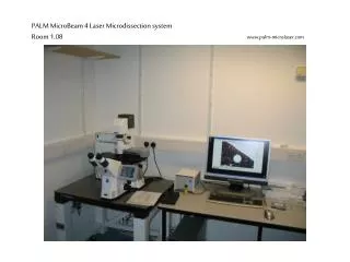
Laser Microdissection Advanced LMD Forensic Applications Patrick Wojtkiewicz, Ph.D. North Louisiana Crime Lab (318) 227-2889 pwojtkie@NLCL.org
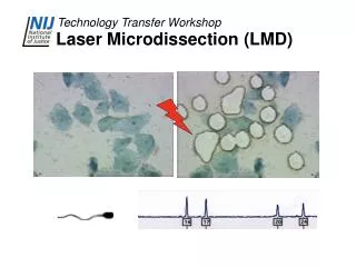
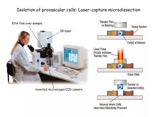
LMD Strengths • Microscopic identification of cells increases sensitivity & minimizes handling • Easily separates Sperm and Epithelial DNA • Quantification is done by cell counting • Fluorescent attachment provides new capabilitie
Fluorescent Capabilities • Adds Capabilities for Analysis of All Types of Sexual Assault Evidence • Provide Improved Capabilities in Sperm Identification • Enable New Capabilities of Identifying Male and Female Diploid Cell Mixtures • These procedures are in the development stage but have been successfully performed on actual case evidence (non-probative)!
Sperm Identification Problems • Few spermatozoa • Searches are often labor intensive • Analyst uncertainty about ID • Case processing based upon sperm id • Excessive quantity of epithelial cells • Spermatozoa buried under cells • Combined with few spermatozoa • Low quality microscope • Poor staining
Sperm Labeling by Fluorescent Dyes • Based upon antibodies conjugated to AlexaFluor. • Antibodies are specific to human sperm components. • Easier, faster, and confident searches • Backwards compatible (not LMD) • Adds extra step in analysis • Requires high quality microscope with fluorescence capabilities
Sperm Paint • Antibodies to:. • ESP (equatorial segment) • SP-10 (acrosomal protein)* • CaBYR-A (tail) • Procedure • Add antibodies to smear • Overnight @ 4ºC • Wash with H2O • Examine
Fluorescent Sperm Identification • Advantages • Simple procedure & Sequential processing of samples • Antibodies should not adversely affect DNA analysis • Samples can be examined at lower magnification • Improved capability - Fast examination & confidence in negative results. Can be done on older slides. • Previous slides are from casework-like material
Problems in Diploid Cell Mixtures • Sample may not be identified as a mixture prior to analysis. • Mixture may not be apparent when there is a preponderance of DNA from one of the donors. • Interpretation issues • No current way of separating diploid cell mixtures.
Chromosome Paint • Fluorescent in situ hybridization (FISH) • X and Y chromosomes • commercial kit • Interphase nuclei • New capability of analyzing male & female epithelial cell mixtures • Time & reproducibility Lymphocytes
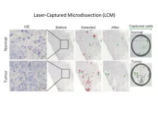
Real Case Results • Identification and dissection of spermatozoa well established • Following results based upon diploid cell analysis • 40% of cases had at least one sample with enough diploid cells to obtain a male profile • Vaginal swab • Bitemark • Fingernail scraping
CODIS Searches In these cases, and other non-probative samples, the profiles were searched locally against approximately 1500 samples. In each case the partial profiles obtained by LMD only hit one sample, the correct one.
Acknowledgements • Jennifer Valentine • Kelli Raley • North Louisiana Crime Lab • LSUSHSC