Download

1 / 12
120 likes | 269 Vues
11.1 Meninges. 2.1 Atoms, Ions, and Molecules. Sponge: Set up Cornell Notes on pg. 15 Topic: 11.1 Meninges Essential Question : 1. NO EQ. BRING BOOKS TOMORROW!!!!!. Pg. 14. Meningitis. Subdural Hematoma. The Central Nervous System (CNS) Brain – lies in the cranial cavity

E N D
11.1 Meninges 2.1 Atoms, Ions, and Molecules Sponge: Set up Cornell Notes on pg. 15 Topic: 11.1 Meninges Essential Question: 1. NO EQ BRING BOOKS TOMORROW!!!!!
Pg. 14 Meningitis Subdural Hematoma
The Central Nervous System (CNS) • Brain – • lies in the cranial cavity • Includes 100 billion multipolar neurons • Brain stem- • connects brain and spinal cord • allows a 2-way communication between brain/spinal cord • Spinal cord- • lies in the vertebral canal within the vertebral column • provides a 2-way communication between the CNS and PNS
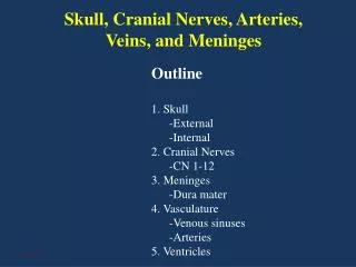
Meninges • Membranes surrounding CNS • Protect the brain and spinal cord • Found between the bone and soft tissue of the n.s. • There are three layers:
Dura mater – • Outermost layer, tough connective tissue • Attaches to the inside of the cranial cavity • Forms the internal covering of the skull bones • Continues into the vertebral canal as a strong tubular covering that surrounds the spinal cord • Contains many blood vessels and nerves • Supports and protects the lobes of the brain and spinal cord
2. Arachnoid mater – • Thin, weblike, Middle layer • Lacks blood vessels • Spreads over the brain and spinal cord • Subarachnoid space lies between arachnoid and piamater which contains the clear watery cerebrospinal fluid or CSF
3. pia mater – • Innermost layer, very thin • many nerves and blood vessels • nourish the underlying cells of the brain and spinal cord
Meninges of the Spinal Cord Subarachnoid space
Disorders Associated with the Meninges: • Meningitis: is an inflammation of the meninges • Caused by Bacteria or viruses that invade the cerebrospinal fluidCommonly limited to the arachnoid and pia maters • Most often in infants and children • Complications: loss of vision, loss of hearing, paralysis and mental retardation. May be FATAL
Subdural hematoma: when a blow to the head ruptures blood vessels associated with the brain • Escaping blood collects between the dura mater and arachnoid mater • Increases pressure between the bones of the skull and the soft tissues of the brain • Will lead to functional losses or even DEATH if not drained
Bottom pg. 14 • Title the page “Video Notes” • 1. Brain Injury • 2. Patrick’s Meningitis • 3. Hand Surgery after Meningitis • 5 bullets each