Understanding Microscopes: A Guide to Light and Electron Microscopes
Microscopes are essential tools for scientists to view and study cells and microscopic structures. The compound light microscope employs a series of lenses to magnify objects. In contrast, the electron microscope, which uses a beam of electrons, magnifies objects up to 1 million times their actual size, revealing atomic arrangements within molecules. Key components include the eyepiece, coarse and fine adjustments, objective lenses, and diaphragms. Proper handling is crucial; always use two hands to carry the microscope to ensure safety.

Understanding Microscopes: A Guide to Light and Electron Microscopes
E N D
Presentation Transcript
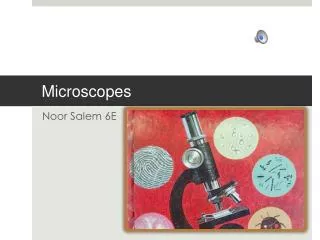
Microscopes Notes Microscopes enabled scientist to view and study the cell.
compound light microscope • It uses series of lenses to magnify objects
Electron Microscope • A new type of microscope was developed called the electron microscope. 1. A beam of electrons instead of light to magnify objects up to 1 million times their actual size. 2. These microscopes can show the arrangement of atoms in a molecule.
1. Ocular lens or eyepiece:10x magnification. • 2. Course Adjustment: Located on side of microscope; innermost is the fine focus and outermost is the coarse focus. • 3. Nosepiece: holds the objective lenses, rotates.
4. Objective lenses: 3 on our scopes, 4x, 10x, 40x Total magnification = eyepiece power x objective power. • 5. Stage: platform on which slides are mounted for viewing • 6. Diaphragm: controls the amount of light which passes to the specimen and can drastically affect the focus of the image.
7. Light: Don’t forget to turn it on or off! • 8. Stage Clip: Holds the slide steady • 9. Base: Keeps the scope from tipping over • 10. Arm: connects the parts of the scope
11.Fine adjustment: small knob on top of big knob. • 12. Body tube: contains mirrors and prisms which direct the image to the ocular lens.
Precaution • ALWAYS use two hands to carry the scope - one on the arm and one under the base - NO EXCEPTIONS!