Download

1 / 77
770 likes | 896 Vues
A retrospective follow-up of ankle fracture patients treated with a biodegradable plate and screws. Background:. Biodegradable fixation implants have been developed to avoid secondary hardware removal.

E N D
A retrospective follow-up of ankle fracture patients treated with a biodegradable plate and screws
Background: Biodegradable fixation implants have been developed to avoid secondary hardware removal.
The aim of this study was to retrospectively follow-up ankle fracture patients treated with a biodegradable plate and screws, and to evaluate the clinical outcome and occurrence of complications.
Methods: Fifty-seven ankle fracture patients treated with biodegradable implants were invited, and a total of 50 were available to participate in this study.
The follow-up included a review of each patient’s medical records, evaluation of radiographs, fracture reduction classification, and functional scoring.
Results: There were 36 lateral malleolar and 14 bimalleolar fractures. No perioperative complications occurred. Average follow-up time was 17 months. All fractures healed.
Fracture alignment was classified as anatomical in 49 patients and good in 1 case. The mean Olerud and Molander functional ankle score at final follow-up was 86.
Eight patients had postoperative complications. These included delayed wound healing in 1 case, 3 cases of deep-vein thrombosis, and 4 soft tissue reactions.
Conclusions: According to the results of this retrospective study, the biodegradable implants used yielded fracture healing and functional results comparable to those previously reported after conventional metal fixation.
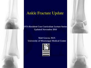
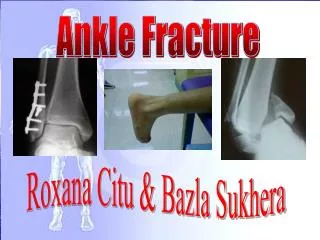
1. Introduction Fractures of the distal fibula (lateral malleolar fractures) are the most common ankle fractures requiring surgical treatment.
Unstable fractures of the lateral malleolus are typically treated with a one-third semitubular or locking metal plate fixed by metal screws to the fibula.
These metal implants provide strong fixation during bone healing but remain in the patient’s body permanently unless subsequently surgically removed.
Although metal implants do not cause any significant problems to most patients, in the USA a secondary procedure for hardware removal after open treatment of lateral malleolar fracture is carried out in 16% of cases during the first postoperative year [1].
Biodegradable materials have been used for years in orthopaedic surgery and traumatology [2,3]. Pins and screws manufactured from these materials have been used with good results in the treatment of ankle fractures [3–11.
Biodegradable plates have been successfully used in the treatment of cranio-maxillofacial and mandibular fractures and osteotomies [12–19], and are currently also being used in many countries in Europe in the treatment of ankle fractures.
Although the idea of treating lateral malleolar fractures with a biodegradable plate is justified as it could make plate removal unnecessary, thereby saving health care resources [6,20,21], only limited clinical data is available on its safety and efficacy in the treatment of fractures of weight-bearing bones [22–25].
The aim of this study was to retrospectively follow-up ankle fracture patients treated with a biodegradable plate and screws, and to evaluate clinical outcome and occurrence of complications in these patients.
2. Materials and methods 2.1. Subjects After ethical committee approval 57 ankle fracture patients treated with a biodegradable plate and screws (Inion OTPSTM, InionOy, Tampere, Finland) between March 2004 and September 2006 were invited for a follow-up visit at a private outpatient clinic.
2.2. Surgical technique All surgical procedures were carried out by one surgeon. After open reduction, all lateral malleolar fractures were fixed with a biodegradable plate and from six to eight biodegradable 2.8 mm or 3.1 mm screws placed through the plate into the distal fibula(Fig. 1).
Fig. 1. The biodegradable implants used (from left to right): 8-hole plate, 2.8 mm screw, 3.1 mm screw, 4.5 mm cortical screw, 4.5 mm cancellous screws (partially and fully threaded), and 2.0 mm pin. The plate can be contoured as desired after 1 min immersion in a hot water bath. The transparency of the plate enables visualisation of the fracture line through the plate during the procedure. The screws and the pins can be cut to the desired length.
In the case of a bimalleolar fracture, the medial malleolus was fixed with biodegradable 4.5 mm cancellous screws and 2.0 mm pins (Inion OTPSTM). Syndesmotic ruptures were treated with a single biodegradable cannulated 4.5 mm cortical screw placed through the plate, across the syndesmosis and through all four cortices of fibula and tibia.
Fracture reduction was checked with fluoroscopy in the operating room. Wounds were closed in normal fashion with biodegradable sutures and covered with absorbent soft silicone dressing.
2.3. Postoperative treatment A below-the-knee cast was applied after the operation and the patients were instructed to use crutches and not to put weight on the affected limb during the first two postoperative weeks.
2 weeks after the operation half body weight bearing was allowed. From the beginning of the fifth postoperative week the patients were allowed to gradually start full weight bearing with pain restriction. The cast was removed 6 weeks postoperatively.
2.4. Follow-up evaluation The follow-up included a review of each patient’s medical records for any remarks regarding perioperative or postoperative complications, or additional medical visits between the day of the original procedure and the follow-up date.
Each patient’s preoperative radiographs, X-rays taken the day after the operation, 6 weeks postoperatively and at the follow-up visit were reviewed, and the final fracture reduction achieved was rated as anatomical, good and poor according to the classification described by Cedell [26] (Table 1).
Clinical outcome at follow-up was evaluated using the functional scoring scale of Olerud and Molander (0–100) [27]. In addition, the duration until return to work and normal daily activities were elicited from each patient.
3. Results A total of 50 patients (21 female, 29 male) gave informed consent to participate in the study. There were 36 lateral malleolar fractures (2 with syndesmosis rupture) and 14 bimalleolar fractures (3 with syndesmosis rupture).
The average age of the patients was 45 years (SD 14, range 18–65) and average weight was 80 kg (SD 18, range 45–150). No perioperative complications occurred. Average follow-up time was 17 months (SD 6.2, range 7–36).
All fractures healed and final fracture reduction was classified as anatomical in 49 patients and good in 1 case (Fig. 2). The mean Olerud and Molander ankle score was 86 (SD 20, range 15–100).
The patient with the lowest score (15) is on disability pension due to her other previous disabilities (unrelated to the ankle fracture). The mean duration before return to work was 2.8 months (SD 1.3, range 1.5–6), and the mean duration before return to normal daily activities 3.1 months (SD 1.3, range 1.5–6).
Eight patients had postoperative complications. These included one delayed wound healing (Fig. 3), 3 cases of deep-vein thrombosis (diagnosed by Doppler ultrasound and treated uneventfully), and four subcutaneous implant-degradation related soft tissue reactions.
The subcutaneous soft tissue reactions were noted as swelling at the implantation site 8–18 months postoperatively (Fig. 4).
In 3 cases, the patients felt that the swelling was minor and painless, they did not complain and would not have consulted a doctor on their own initiative but the reactions were observed by the researchers at the follow-up visit.
In all 4 cases, the swollen area was surgically explored under spinal anesthesia. The plate was observed to have degraded by 18 months but some screw head remnants could be identified inside granulomatous tissue in all cases.
When the incision was made, fluid reminiscent of sour milk was found under the skin. The granulomatous tissue and implant remnants were removed, and samples were collected for microbiological and histopathological evaluation.
The results revealed that no bacterial infection was present but macrophages and giant cells seen microscopically suggested a non-specific foreign body reaction. .
After surgical debridement the wounds were closed in normal fashion and healed uneventfully. The subcutaneous soft tissue reactions observed did not impair wound or bone healing in any of the cases. All fractures had already healed before the reactions occurred
4. Discussion and conclusions According to the results of this retrospective study, the biodegradable plating system used, when used in conjunction with postoperative cast immobilization, provides successful healing of lateral malleolar fractures.
This finding clinically verified the previous assumptions based on the biomechanical results of Sjo¨blom et al. [unpublished data] and Va¨a¨na¨nen et al. [28]. Sjo¨blom et al.
showed in their human cadaver study that the biodegradable plate provides initial fixation stability similar to that of conventional metallic fixation implants and Va¨a¨na¨nen et al.
found in their in vitro degradation study that the plate withstands simulated physiological cyclic loading and maintains its initial fixation stability for 12 weeks, suggesting that the biodegradable plating system is suitable for the treatment of lateral malleolar fractures in conjunction with postoperative cast immobilization.
In addition, the biodegradable screws and pins used provided successful healing of the syndesmosis and medial malleolar fractures in our study.
Fig. 2. Radiographs of a bimalleolar fracture preoperatively (A) and postoperatively (B). The biodegradable implants are not visible in X-rays but the screw holes can be identified.
Eitenmu¨ ller et al. previously investigated the suitability of biodegradable poly-L-lactide screws and plates for the treatment of ankle fractures [22]. In their study, 19 ankle fractures were fixed with 3 mm thick plates and 5.5 mm (1) screws made of blockpolymerised PLLA with high molecular weight. Fractures healed within 6 weeks, but 52% of the patients experienced an aseptic soft tissue problem caused by delayed clearance of the degrading polylactide particles.
Prompted by these problems, the authors treated 7 patients with volume reduced 2 mm thick plates and screws with flat heads, and none of the patients experienced any soft tissue reactions. The authors concluded that the use of biodegradable plates and screws is acceptable for the fixation of ankle fractures, and soft tissue inflammatory reactions can be avoided by using implants with reduced volume of biodegradable material [22].
The biodegradable implants used in the present study are made of co-polymers composed of L-lactide, D-lactide and trimethylene carbonate (TMC). The plates are 1.2 mm thick and the 2.8–3.1 mm (1) screws have flat low-profile heads. The co-polymer material has been found in sheep to become soft in 6–12 months and to degrade completely by 24 months without any harmful inflammatory or foreign body reactions [29].
However, in the present study, subcutaneous implant-degradation related soft tissue reactions were found in four patients (three mild and painless, and one more severe reaction, i.e., 2–8% occurrence rate) 8–18 months postoperatively.
We performed surgical debridement in all 4 cases in this study although only the patient with more severe swelling complained and expressed a desire for treatment. It is not known whether the three milder reactions would have cleared up over time even without surgical treatment.
Fig. 3. Delayed wound healing was observed in one patient 6weeks postoperatively when the cast was removed. The biodegradable implants (the plate and a screw head) were visible in the wound. The patient was treated with antibiotics but the implants were not removed. The wound healed in 4 months without further complications.