Download
1 / 62
750 likes | 2.1k Vues
Embryonic Development of the Brain. 3 rd week – ectoderm thickens to form neural plate , which is later flanked by neural folds This neural groove deepens, forming a neural tube by 4 th week—differentiates into the CNS = brain development begins. Embryonic Development of the Brain.

E N D
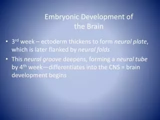
Embryonic Development of the Brain • 3rdweek – ectoderm thickens to form neural plate, which is later flanked by neural folds • This neural groove deepens, forming a neural tube by 4th week—differentiates into the CNS = brain development begins
Embryonic Development of the Brain • Between ectoderm and neural tube a neural crest forms, and the 3 primary brain vesicles appear: 1. FOREBRAIN, or prosencephalon[pros-en-sef-uh-lon] 2. MIDBRAIN, or mesencephalon[mes-en-sef-uh-lon] 3. HINDBRAIN, or rhombencephalon[rom-ben-sef-uh-lon] • The rest of the neural tube becomes the spinal cord
Embryonic Development of the Brain—figure 12.3 • Week 5 – Secondary brain vesicles arise • Forebrain divides: telencephalon[tel-en-sef-uh-lon, -luhn] (“endbrain”) and diencephalon [dahy-en-sef-uh-lon] (“interbrain”) • Hindbrain constricts: metencephalon[met-en-sef-uh-lon] (“afterbrain”) and myelencephalon[mahy-uh-len-sef-uh-lon](“spinal brain”) • Eventually the endbrain sprouts two lateral swellings – like Mickey’s ears • This eventually becomes the cerebrum • The other brain structures form the midbrain, pons, cerebellum and the medulla oblongata [ob-lawng-gah-tuh] • All but the cerebellum form the brain stem
Brain continues to grow rapidly; positions change • Midbrain & cervical flexures develop • Surfaces crease & fold = convolutions • Increase surface area = more neurons
Brain--major parts • Brain stem- continuous with spinal cord • Medulla oblongata, pons, midbrain • Diencephalon [dahy-en-sef-uh-lon] - above brain stem • Thalamus, hypothalamus & pineal gland • Cerebrum- at top and largest part • Surface covered with gray matter- cortex • Beneath is cerebral white matter • Cerebellum- back of brain stem • Means “little brain” • Cranial meninges[mi-nin-jeez]- dura mater, arachnoid[uh-rak-noid] mater & pia mater
Ventricles of the brain-filled with cerebrospinal fluid • Lateral ventricles, one deep within each hemisphere are large C-shaped chambers • They are separated by a thin membrane, the septum pellucidum • Interventricular foramen allows comminucation between the lateral ventricle and the narrow third ventricle • The third and fourth ventricles are connected by the canal-like cerebral aqueduct
The Cerebral Hemisphere • 3 basic regions: • Cerebral cortex • Internal white mater • Basal nuclei (gray matter within white matter) • Surface folds = gyri • Shallow grooves = sulci • Deeper groovesthat separate brain= fissures • Longitudinal Fissure- divides it into left & right hemispheres • Connected by corpus collosum • Transverse fissure- separates cerebral hemispheres from cerebellum
Cerebrum- Structure (cont) • Each hemisphere has 4 lobes • Frontal, parietal, temporal, occipital • Central sulcus separates frontal & parietal • Precentralgyrus anterior to sulcus= primary motor area • Postcentralgyrus = primary sensory area • Each hemisphere is concerned with the sensory and motor functions of the opposite side fo the body. • The two hemispheres are not equal in function.
Function areas of Cortex • Specialized areas anatomically located • Sensory areas receive input and responsible for perception • Motor areas- initiate movements • Associative areas- complex integration: e.g. memory, emotion, reasoning, etc.
Motor Areas • Mainly from anterior part of hemisphere • Primary motor area- precentral gyrus • Broca’s speech area- • interacts with premotor area & primary motor area to regulate breathing and speech muscles
Sensory Areas • Primary somatosensory area-postcentralgyrus. • input includes: touch, proprioception, pain, itching, tickle, temperature • Primary visual area- occipital lobe • Primary auditory area- temporal lobe • Primary gustatory area – base of postcentralgyrus • Primary olfactory area- medial aspect of temporal lobe
Sensory Pathways • Relay information from periphery to cerebral cortex • 3 neurons in each pathway. • Posterior column- medial lemniscus[lem-nis-kuhs]pathway • Fine touch- body location, texture, size • Proprioception-[proh-pree-uh-sep-shuhn] position & motion of body parts • Vibratory sensations- fluctuating touch stimuli
Association Areas • Adjacent to sensory & motor areas • connected with tracts- interpret information • E.g. somatosensory[suh-mat-uh-sen-suh-ree] association area • Posterior to primary somatosensory area • Integrates sensation- exact shape & texture of object compares with stored memories • Wernike’s area- left temporal & parietal [puh-rahy-i-tl]lobes • Interprets meaning of speech • Right hemisphere adds emotional content
Lateralization • Left gets input from & sends output to right side of body and vice versa • Left important for spoken & written language, numerical & scientific skills & reasoning • Right more involved with spatial and pattern recognition and emotional content
Basal Nuclei • Deep gray = basal nuclei (basal ganglia) • Globuspalladus, putamen[pyoo-tey-min], caudate nucleus
Diencephalon • Thalamus- critical relay for sensory input • Transmits motor information from cerebellum & basal nuclei to cerebrum • Hypothalamus- important for homeostasis • Control of ANS-regulation of many activities • Control of pituitary and hormone production • Regulation of emotional & behavior patterns • Regulation of eating & drinking • Control of body temperature • Regulation of circadian rhythms & states of consciousness • Pineal gland- secretes melatonin
Brain Stem- Midbrain • Connects pons to Diencephalon • Large tracts = cerebral peduncles (motor) • Nuclei = substantianigra, rednuclei, cranial nerves III & IV • Superior colliculi – nuclei involved in tracking visual stimuli • Inferior colliculi – auditory input & startle reflex
Brain Stem- Pons • Pons (bridge)- nuclei & tracts • Connect left & right of cerebellum • Ascending & descending tracts • Nuclei – motor relays from cerebrum to cerebellum , respiration & cranial nerves V, VI, VII, VIII
Brain Stem- Medulla • Medulla Oblongata- inferior part of brainstem • white matter extending between spinal cord & other parts of brain • several nuclei: cardiovascular center • (heart rate) • Medullaryrhythmicity area • (respiratory rhythm) • Other sensory & reflex motor areas • Some related to cranial nerves
Cerebellum • Two cerebellar hemispheres • Posterior to medulla and pons, below cerebrum • Cerebellar cortex –gray matter • Tree like white matter & nuclei • Attached to brain stem via cerebellar peduncles
Cerebellar function • Gets wide range of sensory input • Compares with programmed motor activity from cerebral cortex • Smoothes & coordinates complex activities • Regulates posture & balance • Required for skilled motor activities
Limbic System • Ring of structures on inner border of cerebrum and floor of diencephalon • “emotional brain” – pain , pleasure, anger, affection, docility • Involuntary activity related to survival • Important in memory development
Reticular formation • Netlike arrangement of gray and white mater • Ascending part = Reticular Activating System (RAS) • Projects to cerebral cortex & helps maintain consciousness • Inactivation => sleep
Cerebrospinal Fluid (CSF) • Circulates through ventricles of brain and the subarachnoid space. • 4 ventricles: 2 lateral, third & fourth • Formed in choroid plexuses • = Specialized capillary networks in wall of ventricles covered by ependymal cells • Flows through ventricles then from 4th to central canal of spinal cord & subarachnoid cells • Reabsorbed through arachnoid villi into superior saggital sinus
Brain blood supply • Requires ~20% body’s oxygen supply • 4 min lack => permanent damage • Requires continuous glucose supply • Protected by Blood-brain barrier • Allows lipid soluble materials: O2, CO2, alcohol, anesthetic agents but controls entry of other materials • Created by tight capillaries and glial cells
Sensory Pathways (cont) • Spinothalamic pathways- • anterior & lateral spinothalamic tracts • Relay impulses for pain, tickle, itch & thermal sensations.
Somatic Motor Pathways • Signals converge on lower motor neurons • Lower motor neurons stimulate muscles directly • Input comes from: • Local interneurons- e.g. reflexes • Upper motor neurons- corticospinal tracts • Basal ganglia- help with muscle tone • Cerebellum- coordination
Memory • Process for storing & retrieving information • Involves structural & functional changes • Involves association areas, parts of limbic system & diencephalon • Skill memory also involves cerebellum & basal ganglia
Cranial Nerves (table 9.6) • I Olfactory- sensory (smell) • II Optic- sensory (vision) • III Oculomotor- motor (eye) • IV Trochlear- motor (eye) • V Trigeminal- Mixed • Sensory around eyes & upper mouth motor to chewing • VI Abducens- motor (eye) • VII Facial- mixed • Sensory to front of tongue & motor to facial expression, lacrimal and some salivary glands
Cranial Nerves • VIII Vestibulocochlear- sensory (ear) • IX Glossopharyngeal- mixed • Sensory for rest of tongue, pharynx & palate, blood pressure • Motor to pharyngeal muscles, parotid salivary gland • X Vagus- mixed (major visceral nerve) • Sensory from pharynx, ear, diaphragm, visceral organs in ventral cavity • Motor to palatal & pharyngeal muscles & organs in ventral cavity • XI Accessory- Motor to voluntary muscles including sternocleidomastoid and trapezius • XII Hypoglossal- motor to tongue
Aging • Rapid growth during first few years • Size of neurons & proliferation of neuroglia increases • Increases development of dendritic branches & synaptic contacts • Decline in brain mass from early adulthood on
Spinal Cord StructureProtection and Coverings • Spinal cord in vertebral cavity- • Surrounded by bone • Wrapped in meninges- • 3 layers of connective tissue • Spinal cord meninges are continuous with brain meninges
Spinal Meninges • Epidural space lined with fat • Dura mater- tough ,dense connective tissue • Extends to 2nd sacral vertebra • Well beyond spinal cord • Arachnoid mater- collagen and elastic fibers • Subarachnoid space- • cerebral spinal fluid circulates in this space • Pia mater- transparent layer • adheres to surface of brain & spinal cord • Contains blood vessels
Gross Anatomy Of Spinal Cord • Runs to 2nd lumbar vertebra • Roots of spinal nerves for lumbar, sacral & coccygeal nerves in vertebral cavity before leaving = CaudaEquina • Enlargements: cervical & lumbar • Include nerves for upper & lower limbs • Each spinal segment gives rise to a spinal nerve – 31 pairs
Internal Structure Of Spinal Cord • Two grooves- left & right halves • Anterior median fissure & posterior median sulcus • Gray matter- 3 horns on each side • Anterior, posterior, lateral • Anterior- somatic motor neurons • Posterior- sensory neurons • Lateral- autonomic motor neurons