Download
1 / 21
210 likes | 242 Vues
Explore the metabolic, electrophysiological, and circulatory features of the brain and CNS, including neurotransmitter localization, release, and signal transduction. Understand sensory physiology, reflexes, and higher cortical functions. Learn about cerebral blood flow regulation and the resting membrane potential of neurons. Discover the synthesis and receptors of major neurotransmitters and key aspects of somatosensory and pain modulation. Delve into basal ganglia, cerebellum, and cerebral cortex functions along with sleep-wakefulness dynamics and memory mechanisms. Uncover the lateralization of CNS functions and the role of the hypothalamus in regulating bodily functions. Electrophysiology and key neural pathways are also elucidated in this comprehensive guide.

E N D
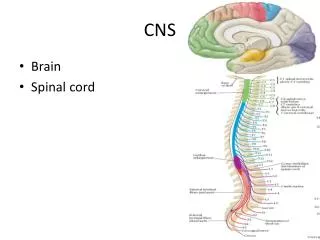
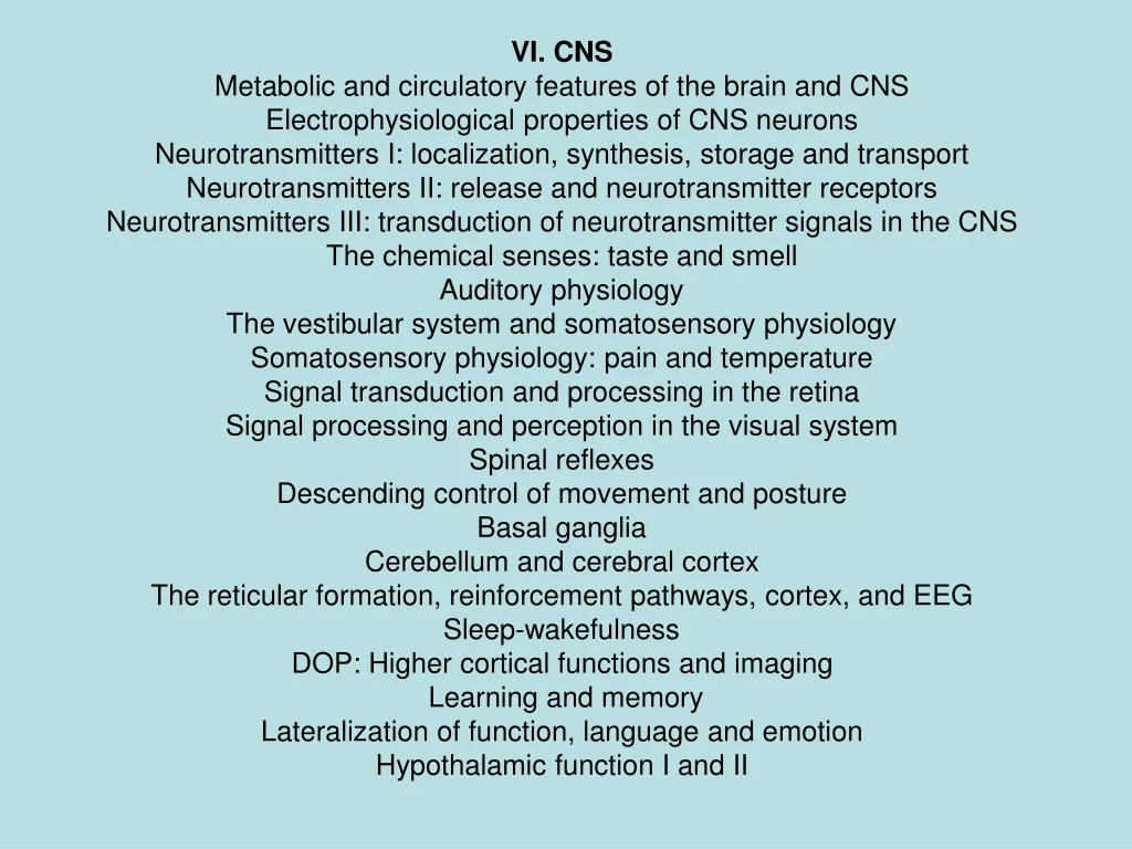
VI. CNS Metabolic and circulatory features of the brain and CNS Electrophysiological properties of CNS neurons Neurotransmitters I: localization, synthesis, storage and transport Neurotransmitters II: release and neurotransmitter receptors Neurotransmitters III: transduction of neurotransmitter signals in the CNS The chemical senses: taste and smell Auditory physiology The vestibular system and somatosensory physiology Somatosensory physiology: pain and temperature Signal transduction and processing in the retina Signal processing and perception in the visual system Spinal reflexes Descending control of movement and posture Basal ganglia Cerebellum and cerebral cortex The reticular formation, reinforcement pathways, cortex, and EEG Sleep-wakefulness DOP: Higher cortical functions and imaging Learning and memory Lateralization of function, language and emotion Hypothalamic function I and II
Brain Metabolism and Circulation 5. Cerebral blood flow increases in cases of a. influenza. b. hypoxia (Arterial P02= 75 mmHg). c. hypertension (Arterjal pressure= 140 mm Hg). d. coma. e. hypercapnia (Arterial PCO2 = 75 mmHg).
Hypercapnia Autoregulation of cerebral blood flow Sympathetic nerve stimulation Regulation of Cerebral Blood Flow 120 Cerebral Blood Flow (ml/min/100g) 60 Remember Metabolic Hyperemia! 60 120 180 Mean Arterial Blood Pressure (mmHg)
Electrophysiology of CNS Neurons The resting membrane potential for all brain neurons is: a. at an equilibrium state for potassium ions. b. at an equilibrium state for sodium ions. c. at an equilibrium state for chloride ions. d. a steady- state level between the equilibrium potentials of the ions involved, weighted by their relative permeabilities/conductances. e. is the same as the resting membrane potential for brain glial cells. Which of the following changes would increase the driving force for sodium entry into a neuron? a. depolarization of the membrane potential b. hyperpolarization of the membrane potential c. an increase in the extracellular sodium concentration d. The sodium equilibrium potential ENa becomes more positive. e. b, c and d are correct.
Membrane Potential • Excitation is caused by: • Depolarization or Hyperpolarization • Increase in gK+ or gNa+ • Increase in ENa or EK
Catecholamine Synthesis AAAD DbH PNMH Tyr Hydroxylase Tyr L-Dopa DA NE Epi MAO or COMT MHPG, VMA, HVA
Neurotransmitter Receptors • GABA • GABAA • Postsynaptic Cl- channel • Agonists- Muscimol, Barbs, (Benzos) • GABAB • G-protein-coupled, activates Adenylate Cyclase • Axoaxonal inhibition. Prevents NT release Pre - GABA GABA GABAB GABA GABA NE GABA Post (-) - GABAA
Neurotransmitter Receptors • Ach • Nicotinic • Cation Channel (Ca++ in CNS) • Spinal Cord, Sup. Colliculus • Muscarinic • G-protein-coupled, Gi inactivates Adenylate Cyclase, Gq activates PLC- Ca++ influx • M1- striatum, hippocampus, cerebrum • M2- cerebellum
Somatosensory • Mechanoreceptors (Ab afferents) • Touch and Pressure (Slow Adapting) • Merkel’s Disk • Ruffini Corpuscles • Touch (Fast Adapting) • Meissner’s (low-freq.) • Hair Follicle • Pacinian Corp. (high-freq.)- wide receptive field • Proprioception • Joint Receptors • Dorsal Column-Medial Lemniscus
Pain Modulation • Nociceptive Fibers (Ad or C)synapse on SC neurons in the anterior horn (A). • Descending 5-HT or NE neurons can modulate these synapses to prevent pain transmission.
Temperature Sense • Cold (Ad) and Warm (C) fibers: • Enter the Spinal Cord at: • Dorsolateral Lissauer fasciculus • Ascend and Descend • Cross SC through ventral w.c. to: • contralateral ALS • Ascend through ALS to reticular formation, thalamus
Axonal Pain Reflex Paper P P (Also bradykinin) Rubor- Red from enlarged arterioles (oxygenated blood) Dolor – Nociception Tumor – Swelling from extravasated fluid P P Type C
Basal Ganglia Disturbances • Parkinsonism • Degradation of DAergic neurons in SN • Tremor, akinesia, bradykinesia, cogwheel rigidity • Huntington’s Chorea • Loss of intrastriatal med. Spiny GABAergics (caudate atrophy) • Chorea, dementia, deceased tone • Ballism • Damage to STN (corpus luysii) - unilateral- hemiballismus • Flailing movements • Athetosis • Damage to GP and Putamen • Wormlike, writhing movements, dystonia (posture issues) • Tardive Dyskinesia • Iatrogenic side effect of neuroleptics (thorazine) • Involuntary mouth movements due to supersensitivity of DA receptors
Cerebellum • Architecture (covered before) • Remember that climbing fibers from inferior olivary nuclei cause complex purkinje spikes, • Mossy fibers from other nuclei cause simple spikes in purkinje cells. • Divisions • Cerebrocerebellum • Spinocerebellum • Vestibulocerebellum • Deep nuclei (send out transmissions) • Fastigial, Interposed (globose,emboliform), Dentate, Lateral Vestibular Nucleus • Granule Cells are the only excitatory ones! – Parallel fibers • Cerebellar Lesions May Cause: • Motor Delay • Dysmetria (inaccuracy) • Dysdiadochokinesia (alternating movement disorders) • Intention Tremor, Ataxia, and Apraxia
Decorticate and DecerebratePostures • Decorticate: CNS Damage above level of Red Nucleus • Rubrospinal Tract active- activates arm flexors with response to pain or head turn in contralateral direction • Decerebrate: CNS Damage at or below level of Red Nucleus • Everything extended except fingers.
The Human Retina • Light changes 11-cis retinal to: • All-trans retinal • This activates: • Transducin • This activates • Phosphodiesterase • This changes: • cGMP to GMP • This causes: • Cation channels to close - hyperpolarization
Sleep Stages • REM- • Sawtooth waves, mixed frequency EEG • Dreaming, Paralysis • NREM- • Stage 1 – Low voltage, mixed freq • Stage 2 – Sleep spindles, K complexes • Stage 3 – Delta waves • Stage 4 – More Delta waves
Reticular Formation • Raphe Nuclei • Serotonin • SN, Ventral Tegmental Area • Dopamine • Nigrostriatal pathway – Motor Control • VTA-frontal cortex and VTA-nucleus accumbens • Dopaminergic neurons overactive in schizophrenia • Also important in reward effects of food, water, and drug abuse • Locus Ceruleus: • Noradrenergic neurons control arousal and sleep-wake cycle
Learning and Memory • AMPA vs. NMDA • AMPA Glu receptors are active during low-frequency stimulation –Na+ channels • NMDA channels require previous depolarization through AMPA channels, allow Ca++ in.