
DEFINITION
DEFINITION. It is the soft tissue covering the Norma Verticalis ( vault of the skull). EXTENSION. It extends from the superciliary arches anteriorly to the external occipital protuberance posteriorly. Laterally , it is continuous to the zygomatic arch. LAYERS.

DEFINITION
E N D
Presentation Transcript
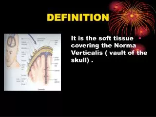
DEFINITION • It is the soft tissue covering the Norma Verticalis ( vault of the skull) .
EXTENSION • It extends from the superciliary arches anteriorly to the external occipital protuberance posteriorly. • Laterally , it is continuous to the zygomatic arch.
LAYERS • The scalp is formed of (Five) layers. • They can be defined by the word itself: • S –Skin. • C –Connective tissue. • A –Aponeurotic layer.
LAYERS • L –Loose connective tissue. • P - Periosteum
SCALP PROPER • It is the first three layers that are tightly held together to form a single unit. • It is the tissue torn away during serious scalping injuries.
SKIN • It is thick hairy with numerous sebaceous and sweat glands. • Obstruction of the ducts of the sebaceous glands by secretions form Sebaceous cysts. • They move with the scalp.
CONNECTIVE TISSUE • It is a fibro-fatty layer which is adherent to the skin and to the underlying aponeurosis by fibrous septa. • It is richly supplied with vessels and nerves embedded within it.
APONEUROTIC LAYER • It is a thin and tendinous sheet that unites the frontal and occipital bellies of occipitofrontalismuscle. • It is attached laterally to the temporal fascia.
OCCIPTOFRONTALIS MUSCLE • It has a frontal belly anteriorly, • An occipital belly posteriorly, and an aponeurotic tendon (galea aponeurotica) connecting the two bellies.
FRONTAL BELLIY • It arises from the anterior part of the aponeurosis. • It is inserted into the skin of the eye brows.
FRONTAL BELLY • It elevates the eyebrows giving the face a surprised looking and produces transverse wrinkles of the forehead.
OCCIPTAL BELLY • It arises from the highest nuchal lines on the occipital bone. • It passes superiorly to be • inserted into the aponeurosis.
NERVE SUPPLY • It is through the terminal branches of the Facial nerve. • The frontal belly is supplied by the temporal branch. • The occipital belly is supplied by the posterior auricular branch.
LOOSE AREOLAR TISSUE • It occupies the subaponeurotic space. • It contains few arteries and the important emissary veins.
DANGEROUS LAYER • The (4th ) layer of the scalp is the dangerous layer because pus or blood spreads easily in it. • Infection in this layer can spreads into the bones through the diploic veins causing osteomyelitis
SCALPINFECTIONS • It can spread through the emissary veins to the intracranial venous sinuses to cause Venous Sinus thrombosis.
SCALP INFECTIONS • An infection in the scalp can not extend posteriorly into the neck because of the attachment of occipitalis muscle to the occipital and temporal bones.
SCALP INFECTIONS • Nor laterally because of attachment of the aponeurosis to the temporal fascia.
SCALP INFECTIONS • An infection or fluid can spreads only into the eye lids and the root of the nose because of the attachment of the frontalis into the skin and not to the bone.
PERICRANIUM • It is the deepest layer. • It is the periosteum on the outer surface of the calvaria. • At the sutures it becomes continuous with the periosteum on the outer surface of the bones.
SENSORY NERVE SUPPLY • It is from two main sources : • Trigeminal nerve. • Cervical nerves (2ND & 3RD ). • Depending on whether it is anterior or posterior to the ears.
ANTERIOR TO THE EAR • (A) Ophthalmic nerve: • 1.Supratrochlear • It exits from the orbit. • It ascends superiorly to supply the forehead and scalp as far as the midline (vertex).
ANTERIOR TO THE EAR • 2. Supraorbital: • It exits from the orbit through the supraorbital notch. • It passes superiorly to the scalp as far as the vertex.
ANTERIOR TO THE EAR • (B) Maxillary nerve: • 3. Zygomaticotemporal nerve: • It exits through a small foramen in the zygomatic bone. • It supplies a small anterior area of the temple.
ANTERIOR TO THE EAR • (C) Mandibular nerve : • 4. Auriculotemporal nerve: • It passes just anterior to the ear. • It supplies the scalp over the temporal region.
POSTERIOR TO THEEAR • 1. Great auricular • It supplies a small area posterior to the scalp. • 2. Lesser occipital: it supplies the area posterior and superior to the scalp.
POSTERIOR TO THEEAR • 3. Greater occipital (posterior ramus of C 2). • 4. Third occipital (posterior ramus of C 3).
ARTERIALSUPPLY • The scalp has a rich blood supply. • The arteries take origin from: • External carotid artery. • Ophthalmic artery. • The arteries freely anastomose with each other.
OPTHALMIC ARTERY • 1. Supratrochlear. • 2. Supraorbital. • They accompany the corresponding nerves to supply the scalp as far as the vertex.
EXTERNAL CAROTID ARTERY • From the posterior aspect: • 1. Posterior auricular: • It is the smallest branch. • It supplies the scalp posterior to the ear.
EXTERNAL CAROTID ARTERY • 2.Occipital : • It accompanies the greater occipital nerve. • It passes through the musculature of the back • It supplies a large area of the back of the scalp.
EXTERNAL CAROTID ARTERY • 3. Superficial temporal artery: • It is the smaller terminal branch of the external carotid. • It divides into anterior and posterior branches. • It supplies almost the entire lateral aspect of the scalp.
VEINS OF THE SCALP • Supratrochlear & supraorbital veins: • They drain the anterior part of the scalp. • They communicate with the ophthalmic veins in the orbit. • Inferiorly they participate in the formation of the angular vein (upper tributary of the(Facial vein).
VEINS OF THE SCALP • Superficial temporal vein: • It drains the entire lateral area of the scalp. • Inferiorly, it joins the maxillary vein to form the Retromandibular vein.
VEINS OF THE SCALP • Posterior auricular vein: • It drains the area posterior to the ear. • It unites with the posterior division of the retromandibular vein to form the External Jugular vein.
VEINS OF THE SCALP • Occipital vein: • It drains into the suboccipital venous plexus. • The plexus drains into the vertebral veins or the internal jugular vein.
VEINS OF THE SCALP Veins of the scalp are connected to the Diploic veins and to the Intracranial venous sinuses through the valveless Emissary veins.
LYMPH DRAINAGE • Lymph vessels follow the arteries. • From the anterior part and forehead drain into : Submandibularnodes.
LYMPH DRAINAGE • Lateral part (above the ear) to: • Superficial parotid(preauricular). • Lateral part (behind the ear) to: • Mastoid nodes. • Back of the scalp to: occipital nodes.
SCALP LACERATIONS • Wounds of the scalp bleed profusely because of: • 1. The abundant arterial anastomoses.
SCALP LACERATIONS • 2. Arteries do not retract when lacerated because they are held open by the dense connective tissue in layer (2). • Local pressure is the only way to stop bleeding.
SCALP LACERATIONS • Deep scalp wounds needs to be sutured because they gape widely when the epicranial aponeurosis is divided. • This because of the tension of the aponeurosis produced by the tone of the occipitofrontalis muscle.